Exploration of the spinal cord and meninges in situ
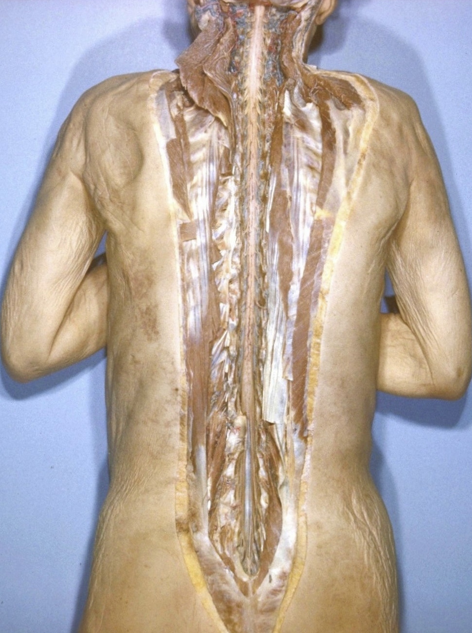
General view of spinal cord; meninges removed
Stanford holds the copyright to the David L. Bassett anatomical images and has assigned
Creative Commons license Attribution-Share
Alike 4.0 International to all of the images.
For additional information regarding use and permissions,
please contact Dr. Drew Bourn at dbourn@stanford.edu.
Image #32-3
Exploration of the spinal cord and meninges in situ
General view of spinal cord; meninges removed
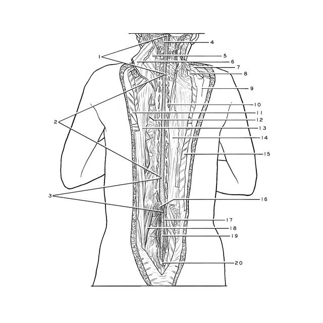
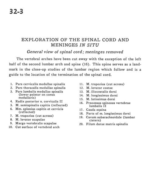
The vertebral arches have been cut away with the exception of the left half of the second lumbar arch and spine (16). This spine serves as a land-mark in the close-up studies of the lumbar region which follow and is a guide to the location of the termination of the spinal cord.
- Cervical part spinal cord
- Thoracic part spinal cord
- Lumbar part spinal cord (lower pointer on conus medullaris)
- Dorsal root cervical nerve II
- Semispinalis capitis muscle (reflected)
- Splenius capitis and cervicis muscles (reflected)
- Trapezius muscle (cut across)
- Levator scapulae muscle
- Scapular-vertebral margin
- Cut surface of vertebral arch
- Trapezius muscle (cut across)
- Levator costarum muscle
- Dorsal iliocostal muscle
- Longissimus dorsi muscle
- Latissimus dorsi muscle
- Spinous process lumbar vertebra II
- Cauda equina
- Parts of longissimus dorsi muscle
- Subarachnoid space (lumbar cistern)
- Filum of dura mater