X CLOSE

Exploration of the spinal cord and meninges in situ

Orientation view; areas of subsequent close-up views indicated

Stanford holds the copyright to the David L. Bassett anatomical images and has assigned Creative Commons license Attribution-Share Alike 4.0 International to all of the images.
For additional information regarding use and permissions, please contact Dr. Drew Bourn at dbourn@stanford.edu.

Image #32-2
Bassett ImageDiagram Image
Legend Image
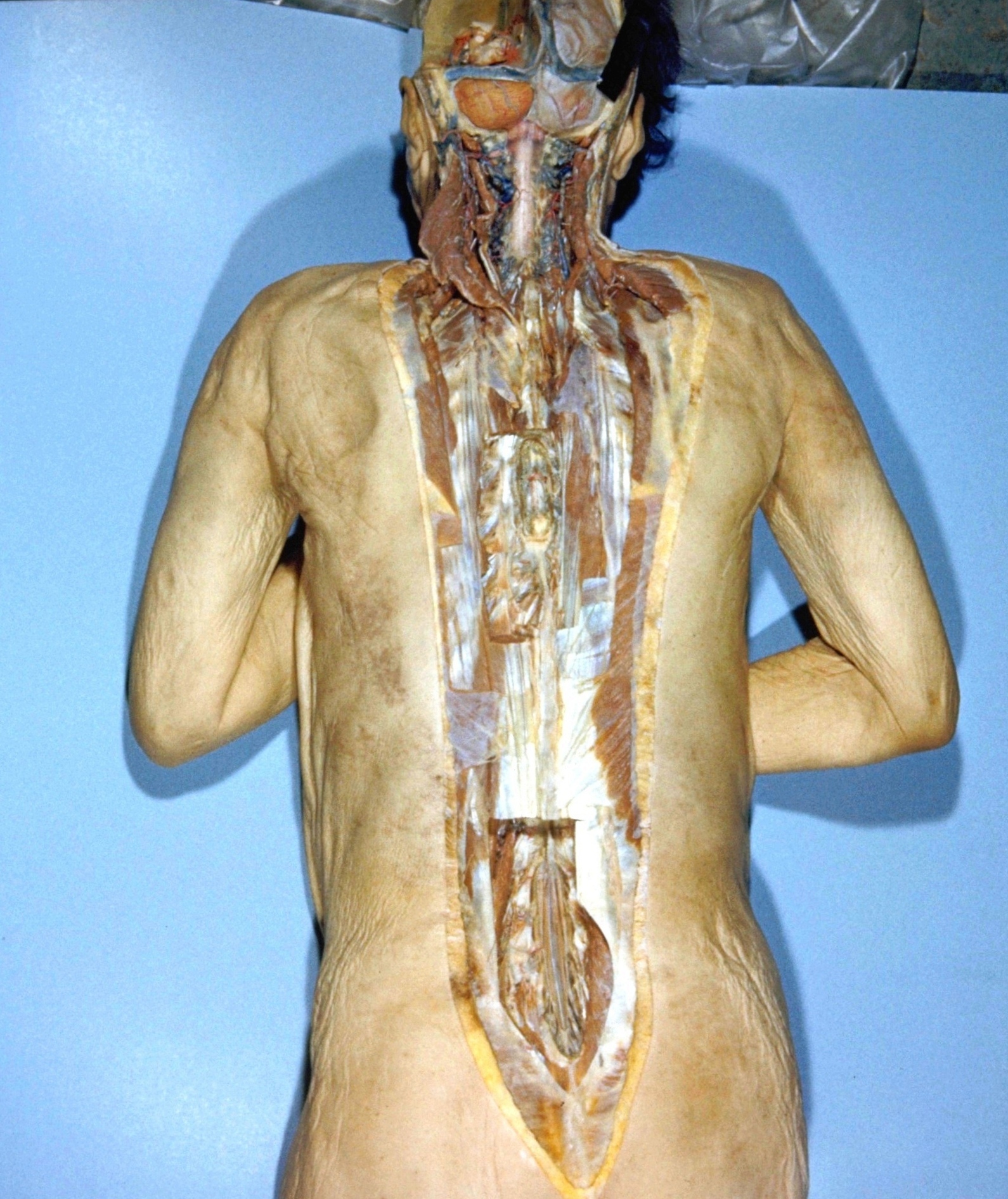
Exploration of the spinal cord and meninges in situ
Orientation view; areas of subsequent close-up views indicated
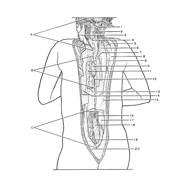
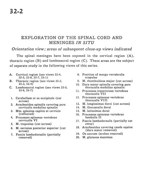
The spinal meninges have been exposed in the cervical region (A), thoracic region (B) and lumbosacral region (C). These areas are the subject of separate study in the following views of this series.
1 . Cerebellum and occipital bone (cut across)
2 . Arachnoid covering cervical part of spinal cord
3 . Splenius capitis and cervicis muscles (reflected)
4 . Spinous process cervical vertebra VI
5 . Trapezius muscle (cut across)
6 . Posterior superior serratus muscle (cut across)
7 . Lumbodorsal fascia (partially removed)
8 . Position of scapular-vertebral margin
9 . Rhomboid major muscle (cut across)
10 . Dura mater covering thoracic part spinal cord
11 . Transverse process thoracic vertebra VII
12 . Spinous process thoracic vertebra VIII
13 . Longissimus dorsi muscle (cut across)
14 . Dorsal iliocostal muscle
15 . Latissimus dorsi muscle
16 . Spinous process lumbar vertebra II
17 . Lumbodorsal fascia (partially cut away)
18 . Arachnoid covering cauda equina (dura mater removed)
19 . Sacrum (arches resected)
20 . Gluteus maximus muscle