Serial transverse sections of the brain stem
Diencephalon.
Stanford holds the copyright to the David L. Bassett anatomical images and has assigned
Creative Commons license Attribution-Share
Alike 4.0 International to all of the images.
For additional information regarding use and permissions,
please contact Dr. Drew Bourn at dbourn@stanford.edu.
Image #31-2
Serial transverse sections of the brain stem
Diencephalon.
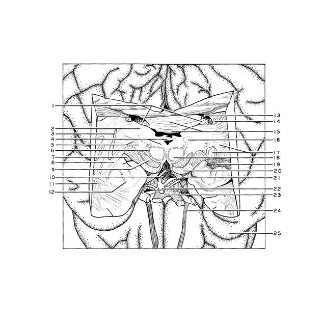
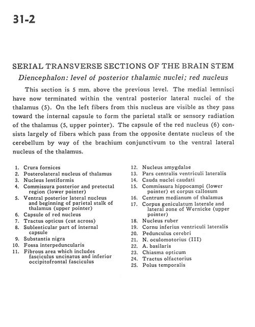
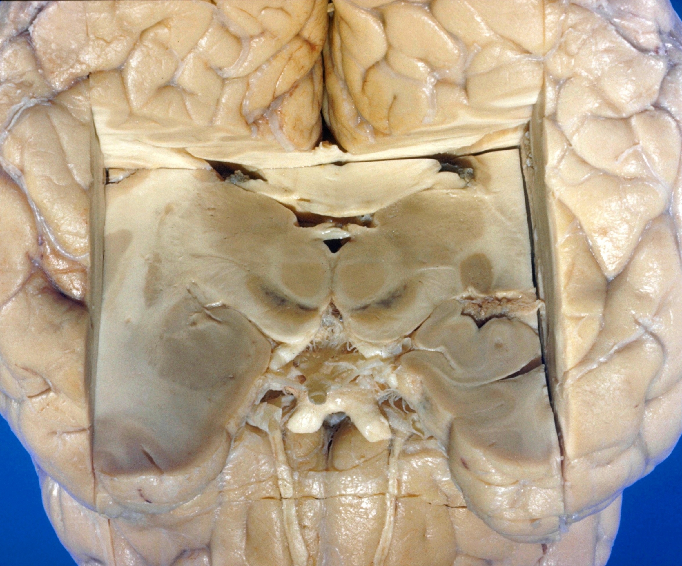
This section is 5 mm. above the previous level. The medial lemnisci have now terminated within the ventral posterior lateral nuclei of the thalamus (5). On the left fibers from this nucleus are visible as they pass toward the internal capsule to form the parietal stalk or sensory radiation of the thalamus (5, upper pointer). The capsule of the red nucleus (6) consists largely of fibers which pass from the opposite dentate nucleus of the cerebellum by way of the brachium conjunctivum to the ventral lateral nucleus of the thalamus.
- Crura fornices
- Posterolateral nucleus of thalamus
- Lentiform nucleus
- Posterior commissure and pretectal region (lower pointer)
- Ventral posterior lateral nucleus and beginning of parietal stalk of thalamus (upper pointer)
- Capsule of red nucleus
- Optic tract (cut across)
- Sublenticular part of internal capsule
- Substantia nigra
- Interpeduncular fossa
- Fibrous area which includes uncinate fasciculus and inferior occipitofrontal fasciculus
- Amygdaloid nucleus
- Central part lateral ventricle
- Caudate nucleus (tail)
- Hippocampal commissure (lower pointer) and corpus callosum
- Centrum (median) of thalamus
- Lateral geniculate body and lateral zone of Wernicke (upper pointer)
- Red nucleus
- Inferior horn of lateral ventricle
- Cerebral peduncle
- Oculomotor nerve (III)
- Basilar artery
- Optic chiasm
- Olfactory tract
- Temporal pole