Serial transverse sections of the brain stem
Pons.
Stanford holds the copyright to the David L. Bassett anatomical images and has assigned
Creative Commons license Attribution-Share
Alike 4.0 International to all of the images.
For additional information regarding use and permissions,
please contact Dr. Drew Bourn at dbourn@stanford.edu.
Image #30-3
Serial transverse sections of the brain stem
Pons.
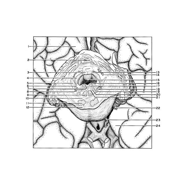
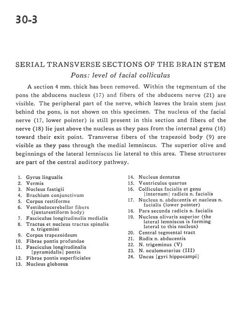
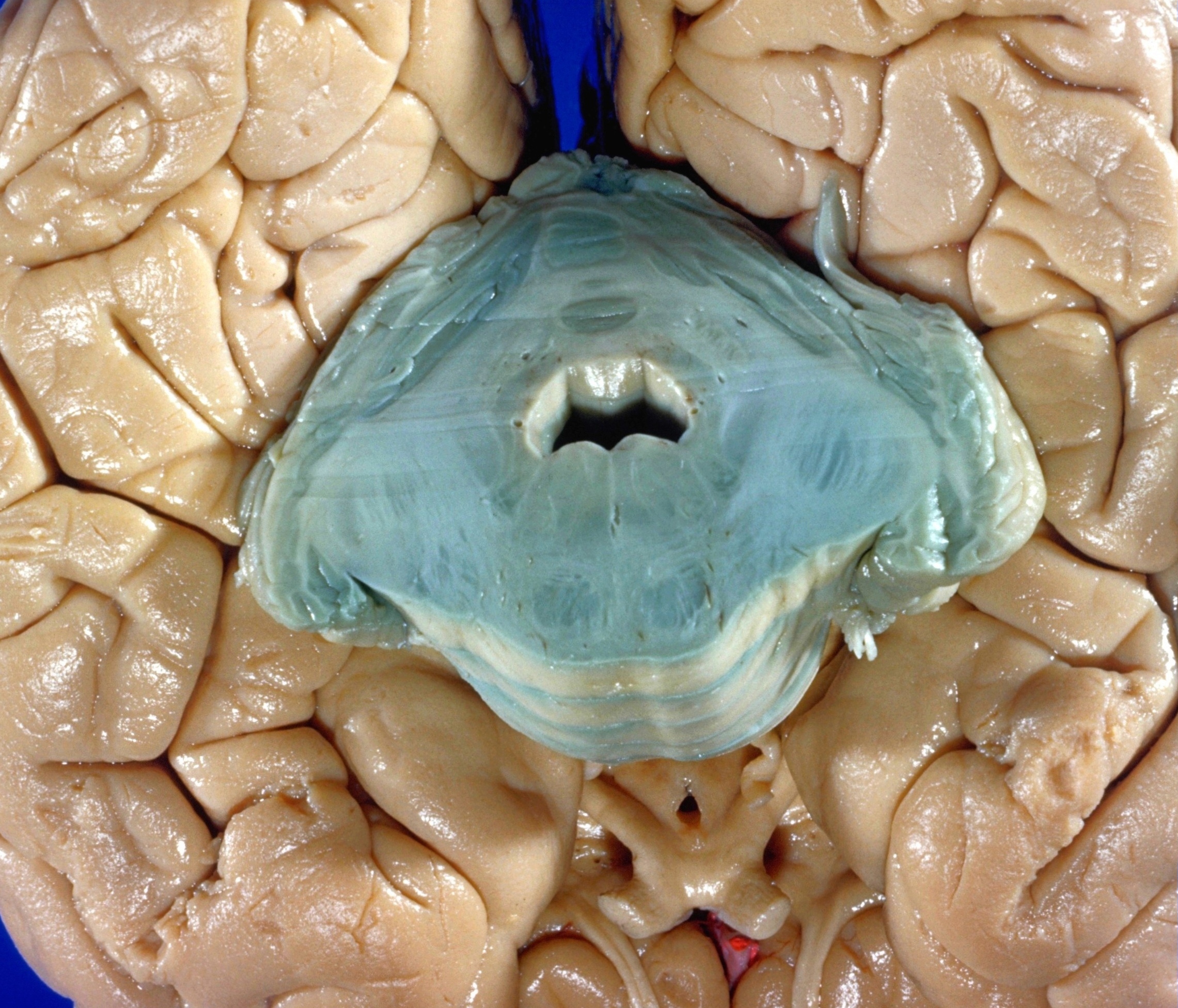
A section 4 mm. thick has been removed. Within the tegmentum of the pons the abducens nucleus (17) and fibers of the abducens nerve (21) are visible. The peripheral part of the nerve, which leaves the brain stem just behind the pons, is not shown on this specimen. The nucleus of the facial nerve (17, lower pointer) is still present in this section and fibers of the nerve (18) lie just above the nucleus as they pass from the internal genu (16) toward their exit point. Transverse fibers of the trapezoid body (9) are visible as they pass through the medial lemniscus. The superior olive and beginnings of the lateral lemniscus lie lateral to this area. These structures are part of the central auditory pathway.
- Lingual gyrus
- Vermis
- Fastigial nucleus
- Brachium conjunctivum (superior cerebellar peduncle)
- Restiform body (inferior cerebellar peduncle)
- Vestibulocerebellar fibers (juxtarestiform body)
- Medial longitudinal fasciculus
- Spinal trigeminal tract and nucleus
- Trapezoid body
- Deep pontine fibers
- Longitudinal fasciculus (pontine pyramid)
- Superficial pontine fibers
- Globose nucleus
- Dentate nucleus
- Fourth ventricle
- Facial colliculus and genu (internal) roots of facial nerve
- Nucleus of abducens nerve and nucleus of facial nerve (lower pointer)
- Second part of roots of facial nerve (VII)
- Superior olivary nucleus (the lateral lemniscus is forming lateral to this nucleus)
- Central tegmental tract
- Root abducens nerve
- Trigeminal nerve (V)
- Oculomotor nerve (III)
- Uncus (hippocampal gyrus)