For permissions information regarding the use of these images,
please contact Drew Bourn at dbourn@stanford.edu
Image #29-4
Serial transverse sections of the brain stem
Medulla oblongata.
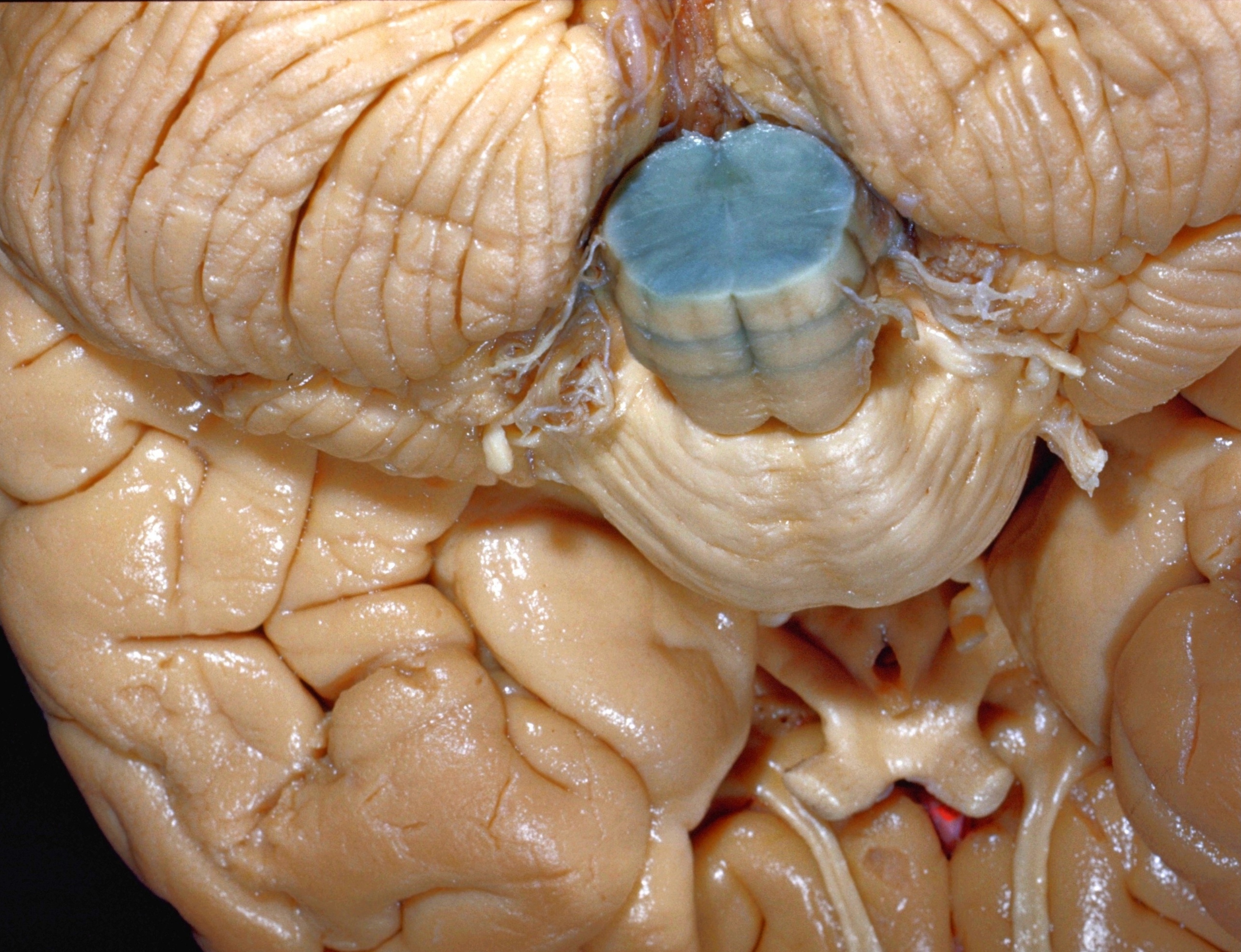
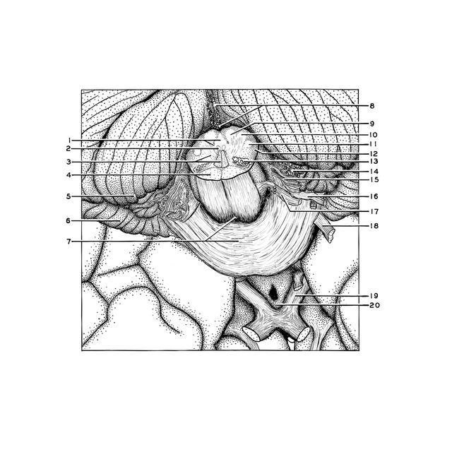
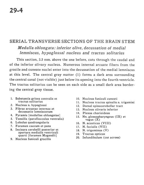
This section, 3.5 mm. above the one before, cuts through the caudal end of the inferior olivary nucleus. Numerous internal arcuate fibers from the gracile and cuneate nuclei enter into the decussation fo the medial lemniscus at this level. The central gray matter (1) forms a dark area surrounding the central canal (not visible) just below its opening into the fourth ventricle. The tractus solitarius can be seen on each side as a small dark area bordering the central gray tissue.
1
.
Central gray matter and tractus solitarius
2
.
Nucleus hypoglossal nerve (XII)
3
.
Internal arcuate fibers and decussation of lemniscus
4
.
Pyramid (medulla oblongata)
5
.
Tonsil (ventral paraflocculus)
6
.
Quadrangular lobule
7
.
Foramen caecum and pons
8
.
Posterior cerebellar incisure and medial aperture fourth ventricle (foramen of magendie)