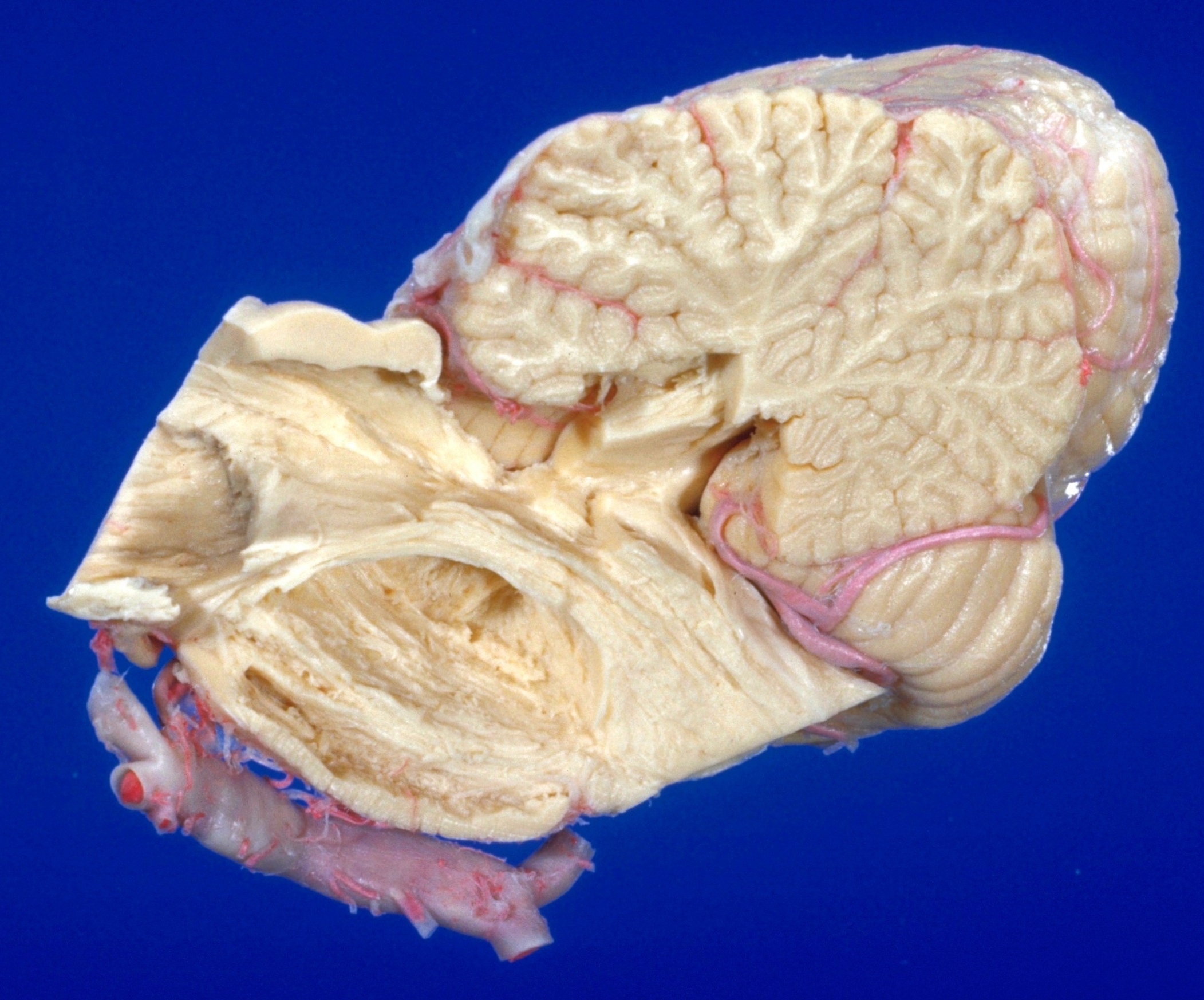
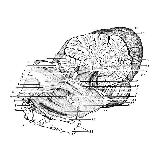
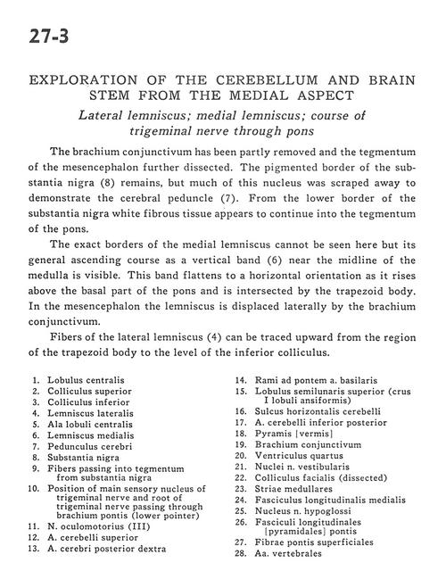
Exploration of the cerebellum and brain stem from the medial aspect
Lateral lemniscus; medial lemniscus; course of trigeminal nerve through pons
Stanford holds the copyright to the David L. Bassett anatomical images and has assigned
Creative Commons license Attribution-Share Alike 4.0 International to all of the images.
For additional information regarding use and permissions,
please contact Dr. Drew Bourn at dbourn@stanford.edu.
Image #27-3
 |  | ||||||||||||||||||||||||||||||||||||||||||||||||||||||||||||
 |
|