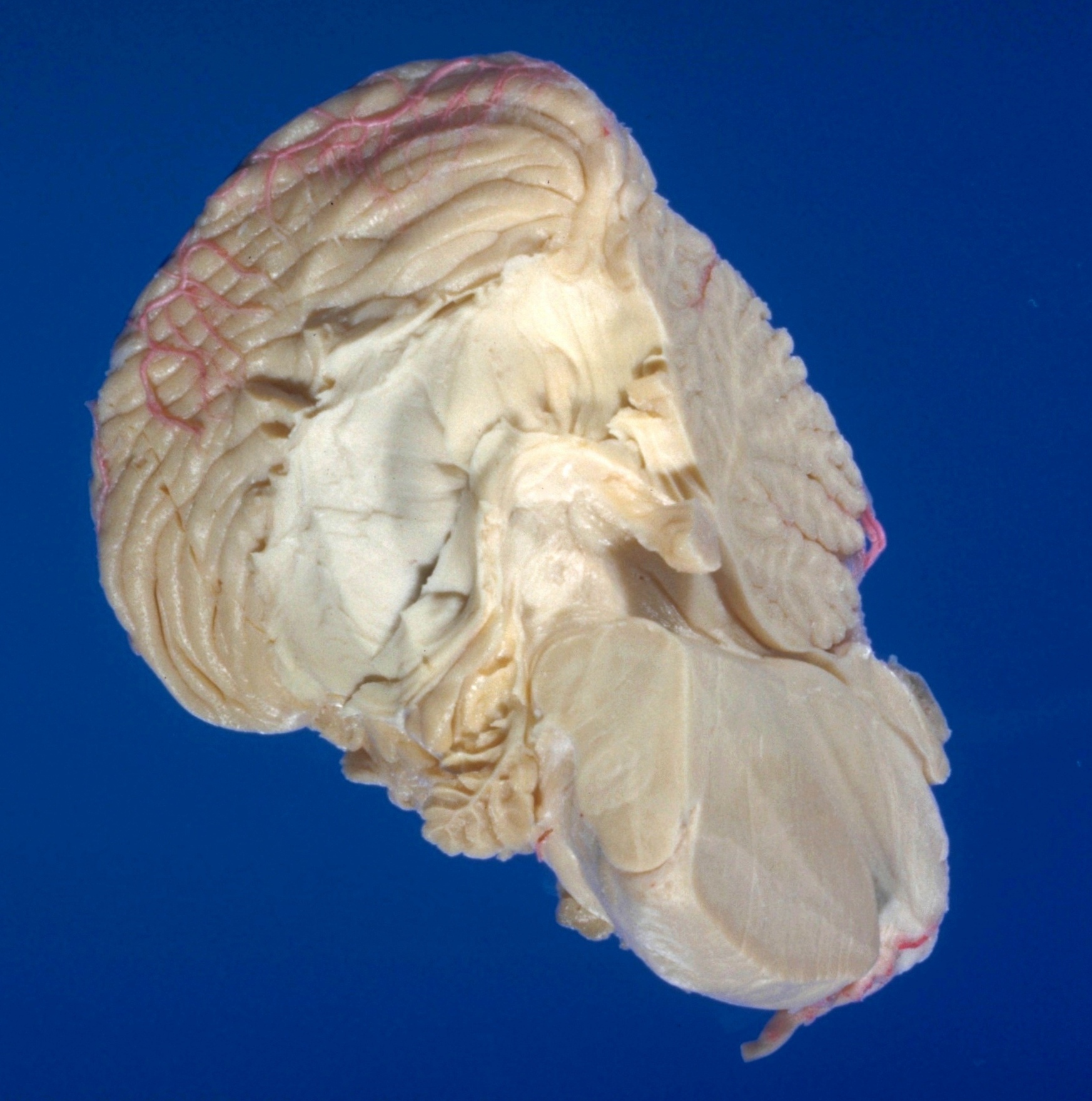
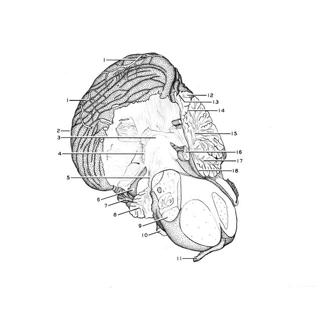
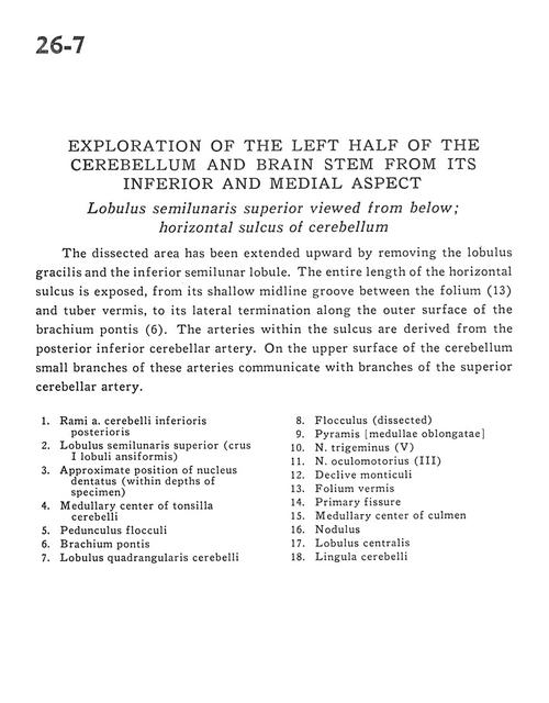
Exploration of the left half of the cerebellum and brain stem from its inferior and medial aspect
Lobulus semilunaris superior viewed from below; horizontal sulcus of cerebellum
Stanford holds the copyright to the David L. Bassett anatomical images and has assigned
Creative Commons license Attribution-Share Alike 4.0 International to all of the images.
For additional information regarding use and permissions,
please contact Dr. Drew Bourn at dbourn@stanford.edu.
Image #26-7
 |  | ||||||||||||||||||||||||||||||||||||||||
 |
|