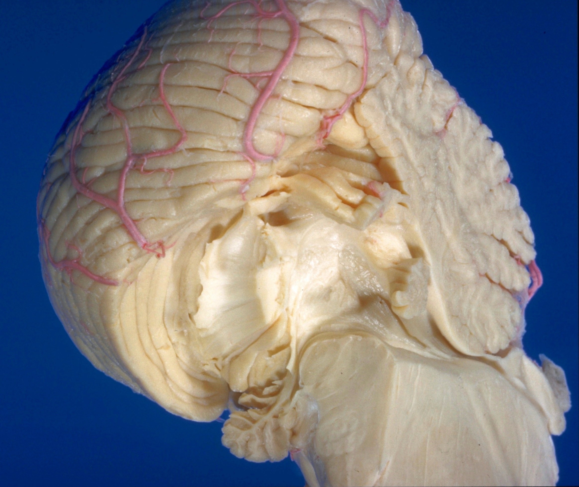
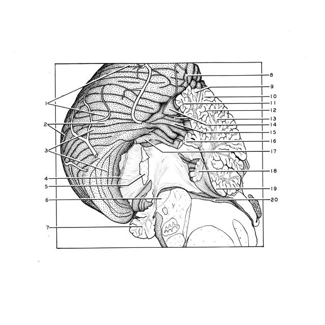
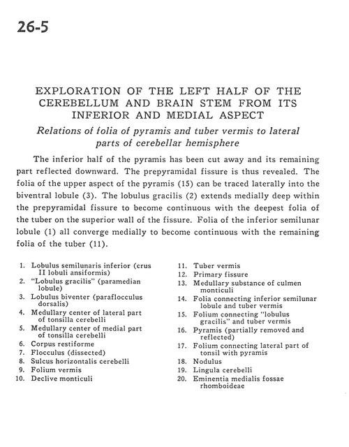
Exploration of the left half of the cerebellum and brain stem from its inferior and medial aspect
Relations of folia of pyramis and tuber vermis to lateral parts of cerebellar hemisphere
Stanford holds the copyright to the David L. Bassett anatomical images and has assigned
Creative Commons license Attribution-Share Alike 4.0 International to all of the images.
For additional information regarding use and permissions,
please contact Dr. Drew Bourn at dbourn@stanford.edu.
Image #26-5
 |  | ||||||||||||||||||||||||||||||||||||||||||||
 |
|