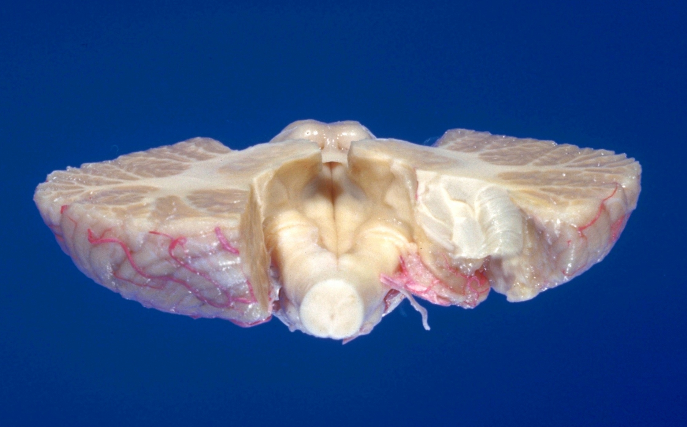
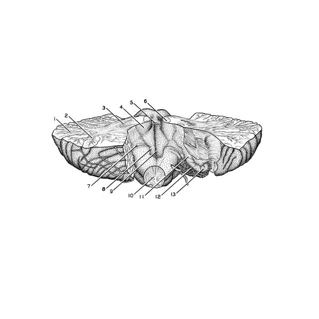
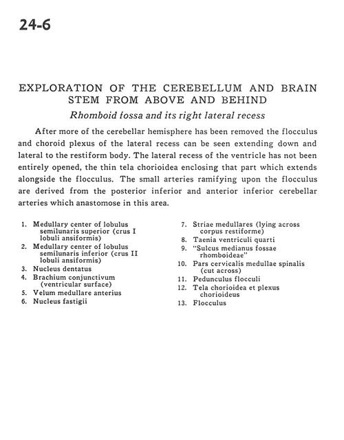
Exploration of the cerebellum and brain stem from above and behind
Rhomboid fossa and its right lateral recess
Stanford holds the copyright to the David L. Bassett anatomical images and has assigned
Creative Commons license Attribution-Share Alike 4.0 International to all of the images.
For additional information regarding use and permissions,
please contact Dr. Drew Bourn at dbourn@stanford.edu.
Image #24-6
 |  | ||||||||||||||||||||||||||||||
 |
|