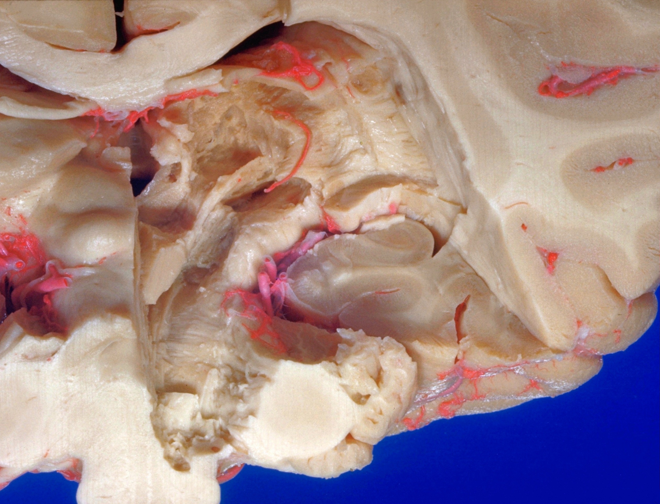
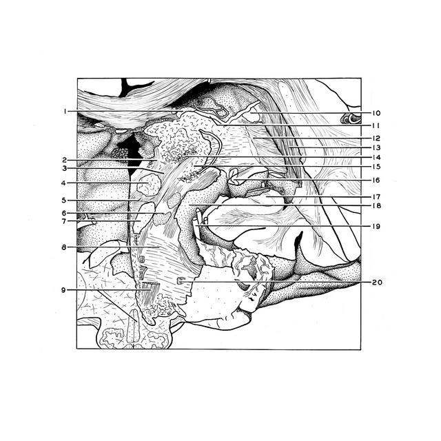
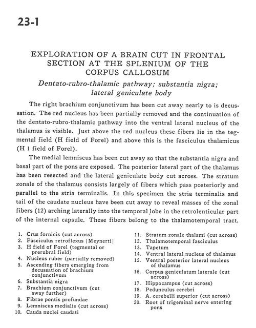
Exploration of a brain cut in frontal section at the splenium of the corpus callosum
Dentato-rubro-thalamic pathway; substantia nigra; lateral geniculate body
Stanford holds the copyright to the David L. Bassett anatomical images and has assigned
Creative Commons license Attribution-Share Alike 4.0 International to all of the images.
For additional information regarding use and permissions,
please contact Dr. Drew Bourn at dbourn@stanford.edu.
Image #23-1
 |  | ||||||||||||||||||||||||||||||||||||||||||||
 |
|