X CLOSE

Dissection of anterior aspect of vertebral column

Coccygeal region.

Stanford holds the copyright to the David L. Bassett anatomical images and has assigned Creative Commons license Attribution-Share Alike 4.0 International to all of the images.
For additional information regarding use and permissions, please contact Dr. Drew Bourn at dbourn@stanford.edu.

Image #221-7
Bassett ImageDiagram Image
Legend Image
Dissection of anterior aspect of vertebral column
Coccygeal region.
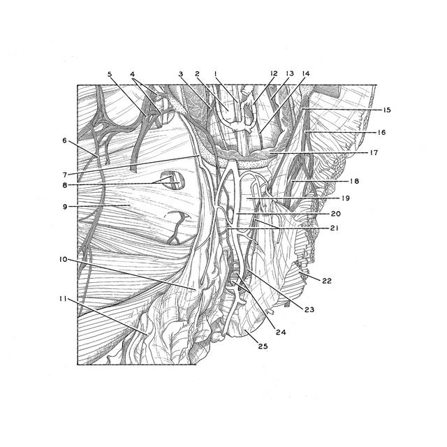
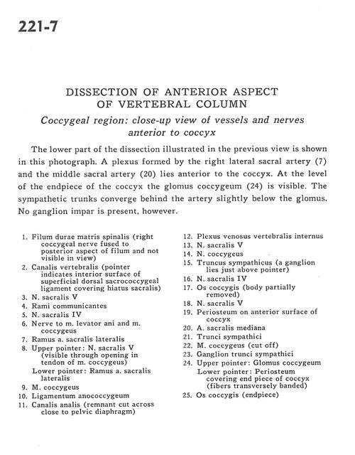
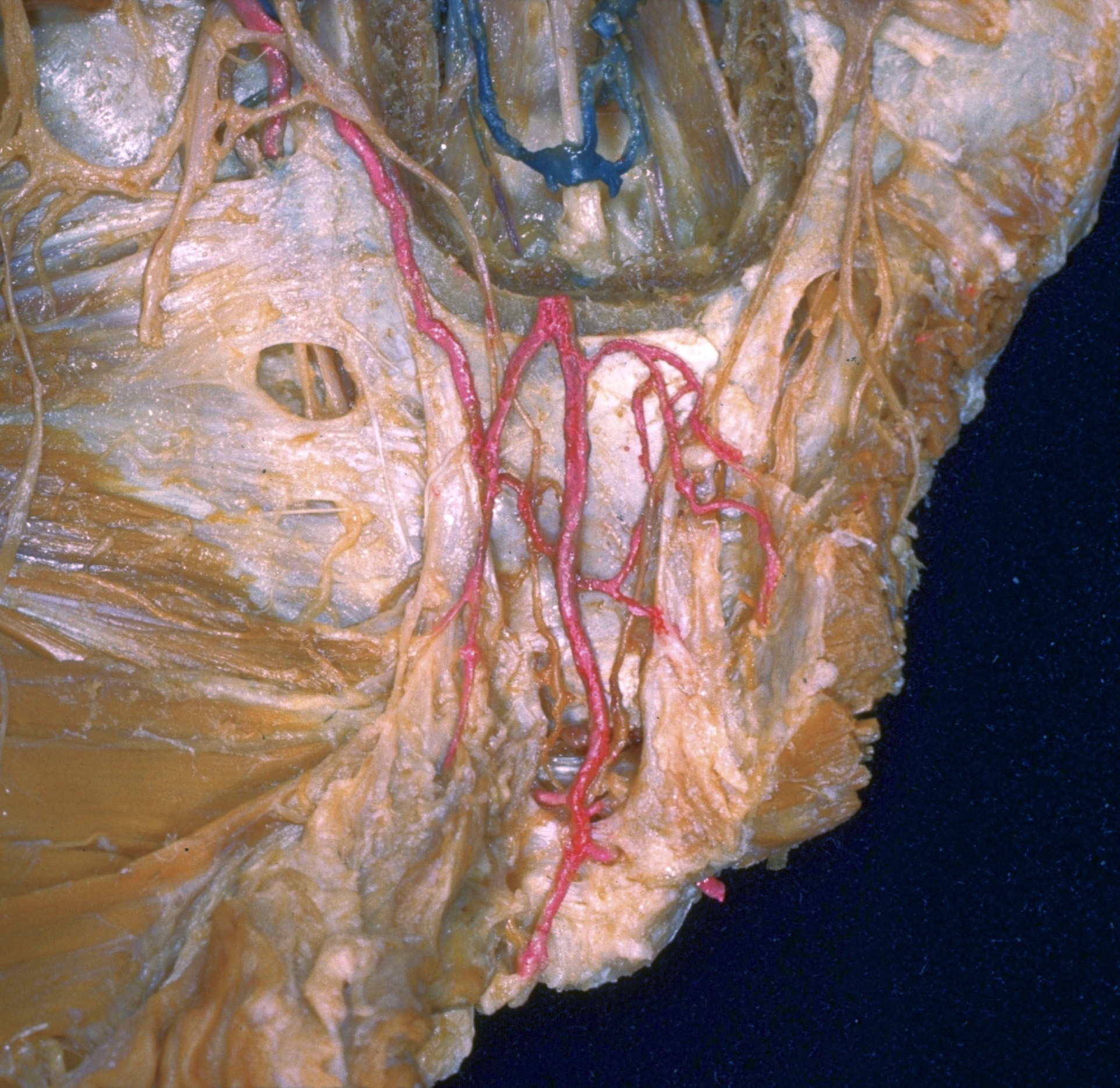
The lower part of the dissection illustrated in the previous view is shown in this photograph. A plexus formed by the right lateral sacral artery (7) and the middle sacral artery (20) lies anterior to the coccyx. At the level of the endpiece of the coccyx the glomus coccygeum (24) is visible. The sympathetic trunks converge behind the artery slightly below the glomus. No ganglion impar is present, however.
1 . Filum terminale (right coccygeal nerve fused to posterior aspect of filum and not visible in view)
2 . Vertebral canal (pointer indicates interior surface of superficial dorsal sacrococcygeal ligament covering sacral hiatus)
3 . Sacral nerve V
4 . Rami communicantes
5 . Sacral nerve IV
6 . Nerve to levator ani muscle and coccygeus muscle
7 . Ramus lateral sacral artery
8 . Upper pointer: Sacral nerve V (visible through opening in tendon of coccygeus muscle) Lower pointer: Lateral branch sacral artery
9 . Coccygeus muscle
10 . Anococcygeal ligament
11 . Anal canal (remnant cut across close to pelvic diaphragm)
12 . Internal vertebral venous plexus
13 . Sacral nerve V
14 . Coccygeal nerve
15 . Sympathetic trunk (a ganglion lies just above pointer)
16 . Sacral nerve IV
17 . Coccyx (body partially removed)
18 . Sacral nerve V
19 . Periosteum on anterior surface of coccyx
20 . Median sacral artery
21 . Sympathetic trunk
22 . Coccygeus muscle (cut off)
23 . Ganglion of sympathetic trunk
24 . Upper pointer: Glomus coccygeum Lower pointer: Periosteum covering end piece of coccyx (fibers transversely banded)
25 . Coccyx (endpiece)