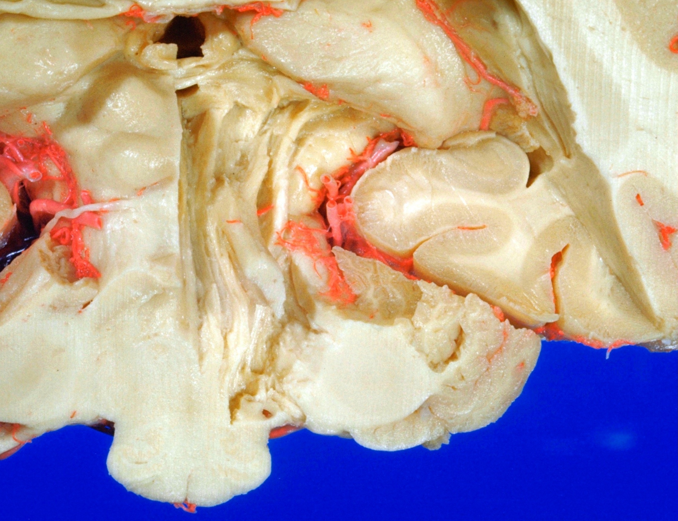
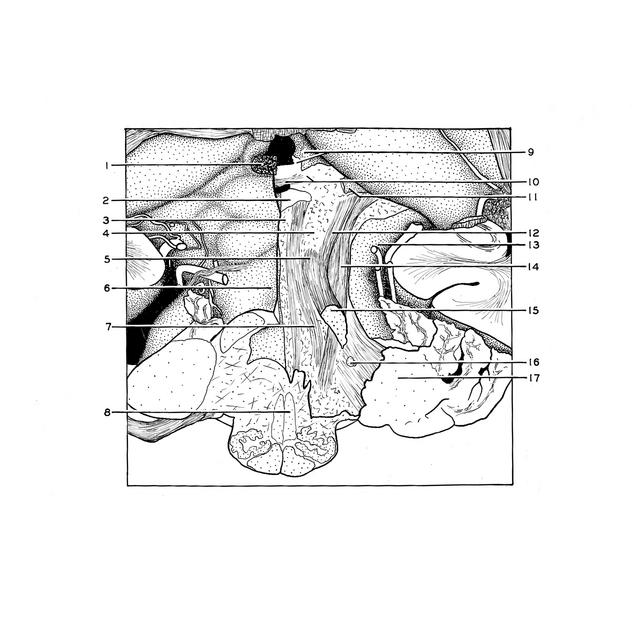
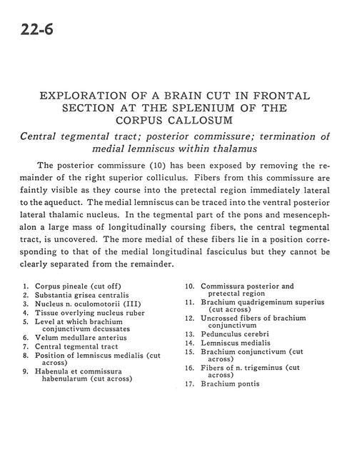
Exploration of a brain cut in frontal section at the splenium of the corpus callosum
Central tegmental tract; posterior commissure; termination of medial lemniscus within thalamus
Stanford holds the copyright to the David L. Bassett anatomical images and has assigned
Creative Commons license Attribution-Share Alike 4.0 International to all of the images.
For additional information regarding use and permissions,
please contact Dr. Drew Bourn at dbourn@stanford.edu.
Image #22-6
 |  | ||||||||||||||||||||||||||||||||||||||
 |
|