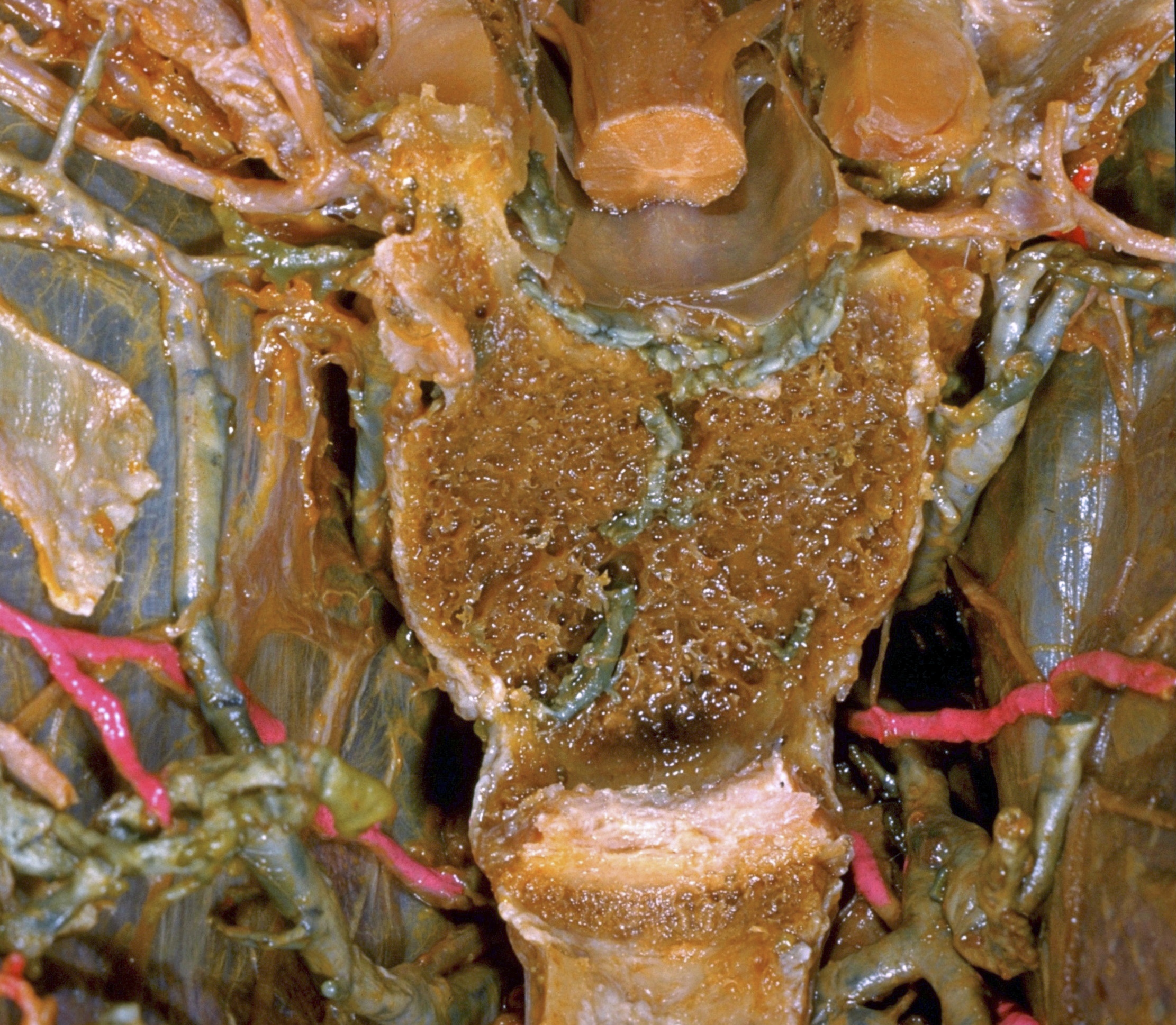
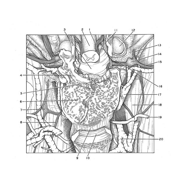
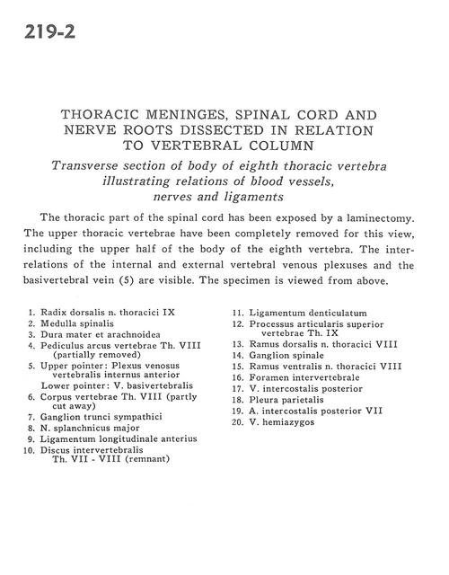
Thoracic meninges, spinal cord and nerve roots dissected in relation to vertebral column
Transverse section of body of eighth thoracic vertebra illustrating relations of blood vessels, nerves and ligaments
Stanford holds the copyright to the David L. Bassett anatomical images and has assigned
Creative Commons license Attribution-Share Alike 4.0 International to all of the images.
For additional information regarding use and permissions,
please contact Dr. Drew Bourn at dbourn@stanford.edu.
Image #219-2
 |  | ||||||||||||||||||||||||||||||||||||||||||||
 |
|