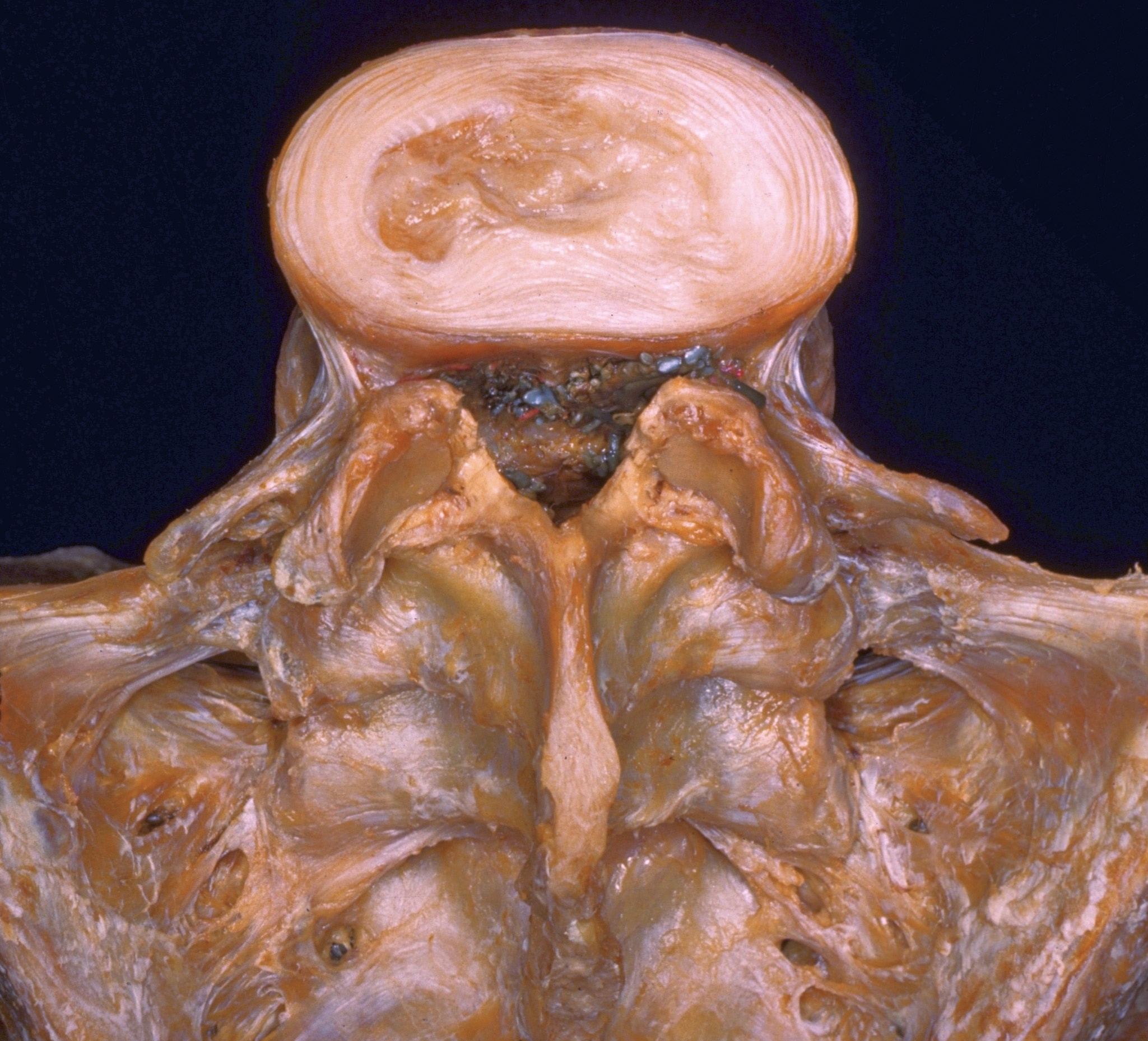
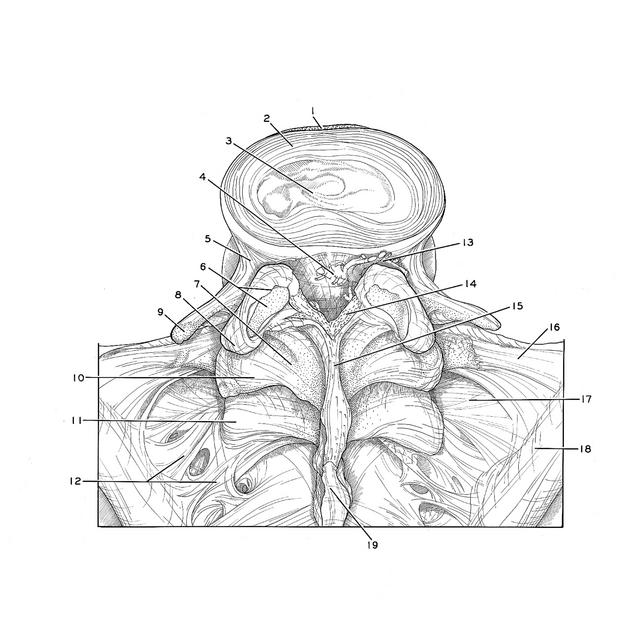
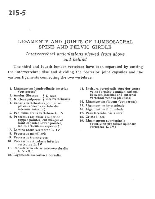
Ligaments and joints of lumbosacral spine and pelvic girdle
Intervertebral articulations viewed from above and behind
Stanford holds the copyright to the David L. Bassett anatomical images and has assigned
Creative Commons license Attribution-Share Alike 4.0 International to all of the images.
For additional information regarding use and permissions,
please contact Dr. Drew Bourn at dbourn@stanford.edu.
Image #215-5
 |  | ||||||||||||||||||||||||||||||||||||||||||
 |
|