X CLOSE

Ligaments and joints of lumbosacral spine and pelvic girdle

Ligaments of sacral region, posterior view

Stanford holds the copyright to the David L. Bassett anatomical images and has assigned Creative Commons license Attribution-Share Alike 4.0 International to all of the images.
For additional information regarding use and permissions, please contact Dr. Drew Bourn at dbourn@stanford.edu.

Image #215-1
Bassett ImageDiagram Image
Legend Image
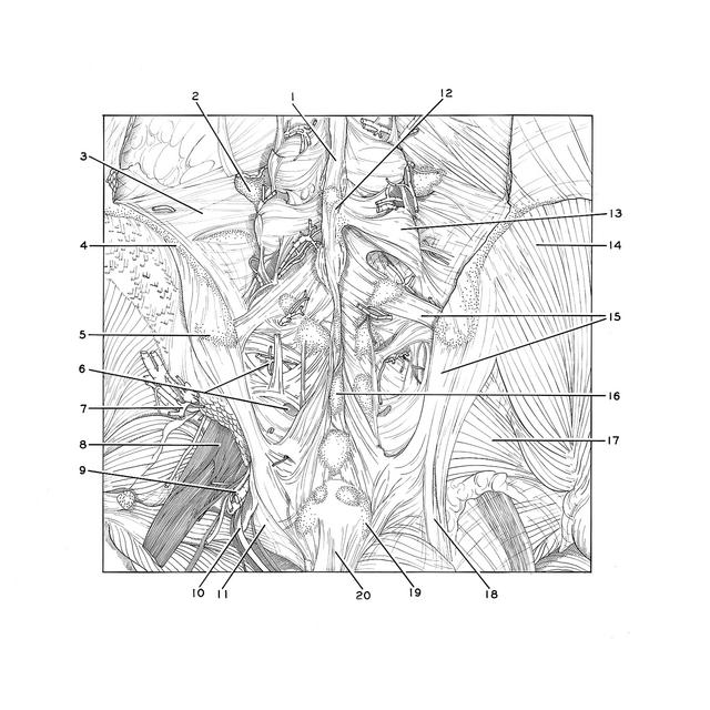
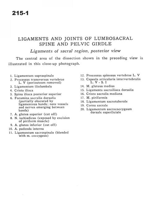
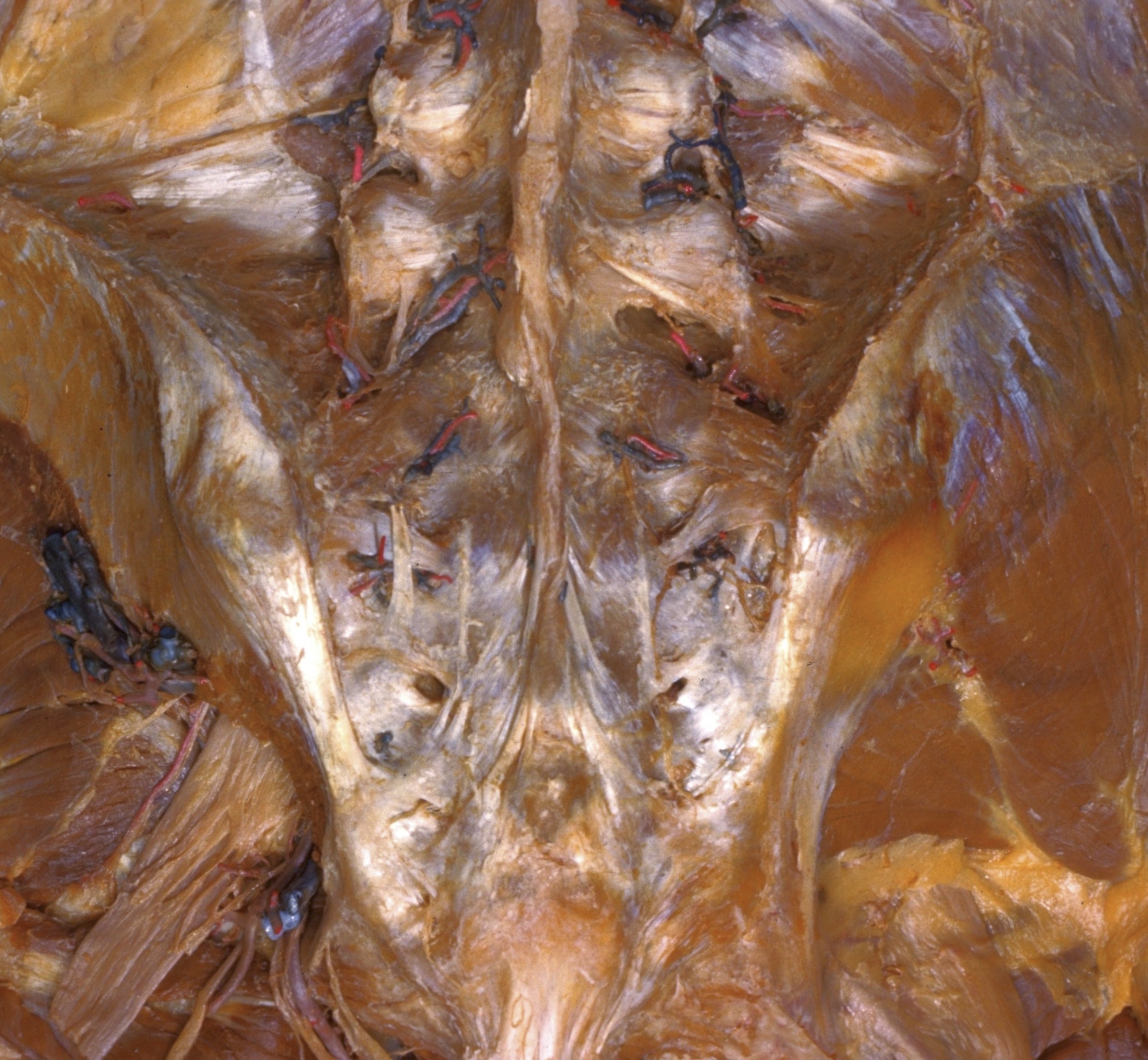
Ligaments and joints of lumbosacral spine and pelvic girdle
Ligaments of sacral region, posterior view
The central area of the dissection shown in the preceding view is illustrated in this close-up photograph.
1 . Supraspinous ligament
2 . Transverse process of vertebra L. V (periosteum removed)
3 . Iliolumbar ligament
4 . Iliac crest
5 . Posterior superior iliac spine
6 . Dorsal sacral foramina (partially obscured by ligamentous bands note vessels and nerves emerging between bands)
7 . Superior gluteal artery (cut off)
8 . Sciatic nerve (exposed by excision of piriform muscle)
9 . Inferior gluteal artery (cut off)
10 . Internal pudendal artery
11 . Sacrospinous ligament (blended with coccygeus m)
12 . Spinous process vertebra L. V
13 . Intervertebral joint capsule L. V - S. I
14 . Gluteus medius muscle
15 . Dorsal sacroiliac ligament
16 . Median sacral crest
17 . Piriform muscle
18 . Sacrotuberous ligament
19 . Sacral cornu (horn)
20 . Dorsal superficial sacrococcygeal ligament