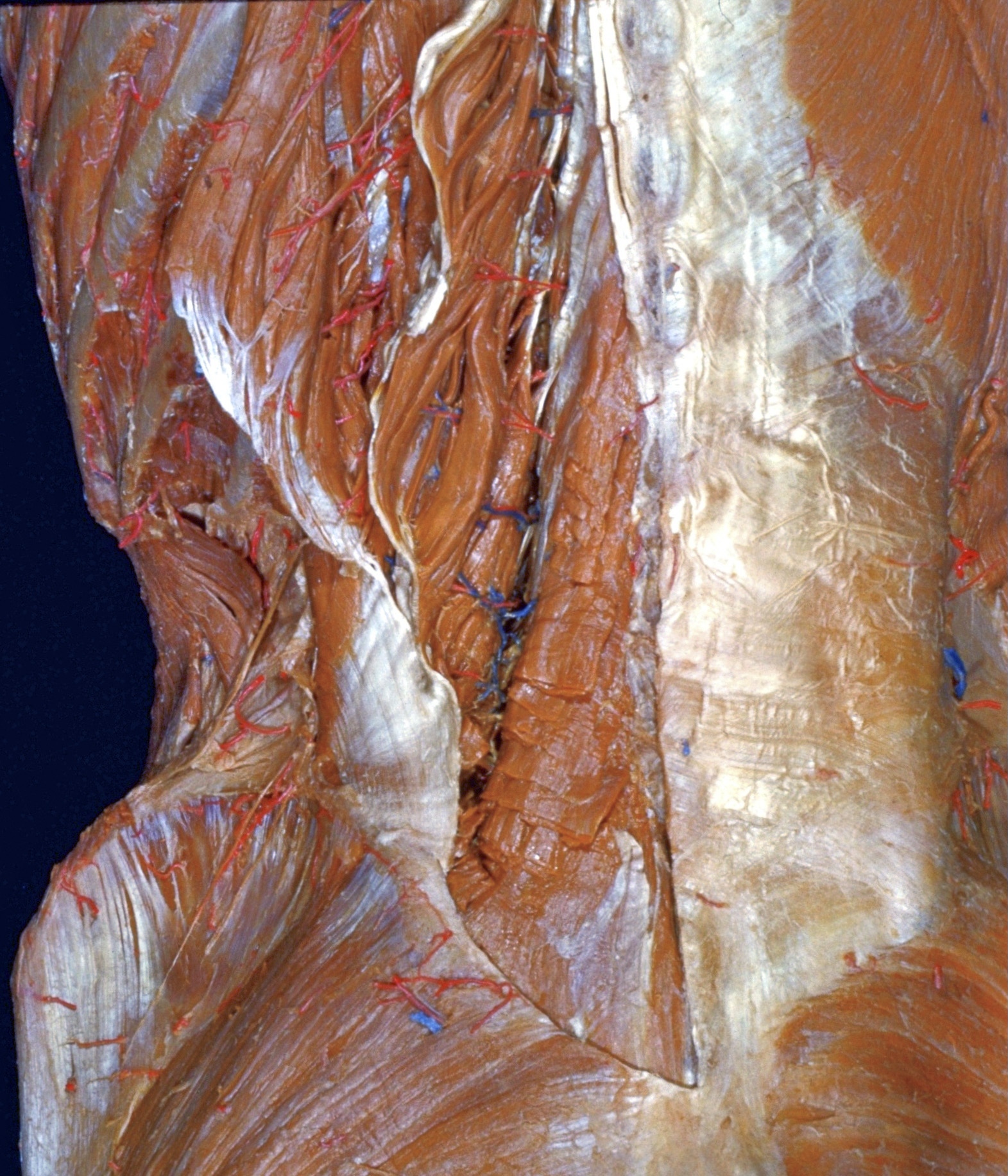
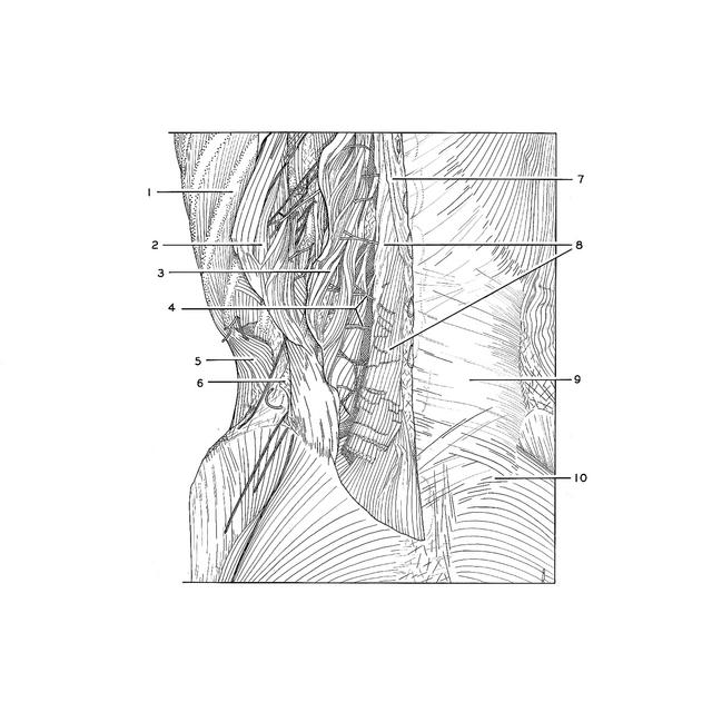
Dissection of thoracic and lumbosacral regions of back from a posterior approach
Multifidus muscle in lumbar region
Stanford holds the copyright to the David L. Bassett anatomical images and has assigned
Creative Commons license Attribution-Share Alike 4.0 International to all of the images.
For additional information regarding use and permissions,
please contact Dr. Drew Bourn at dbourn@stanford.edu.
Image #214-1
 |  | ||||||||||||||||||||||||
 |
|