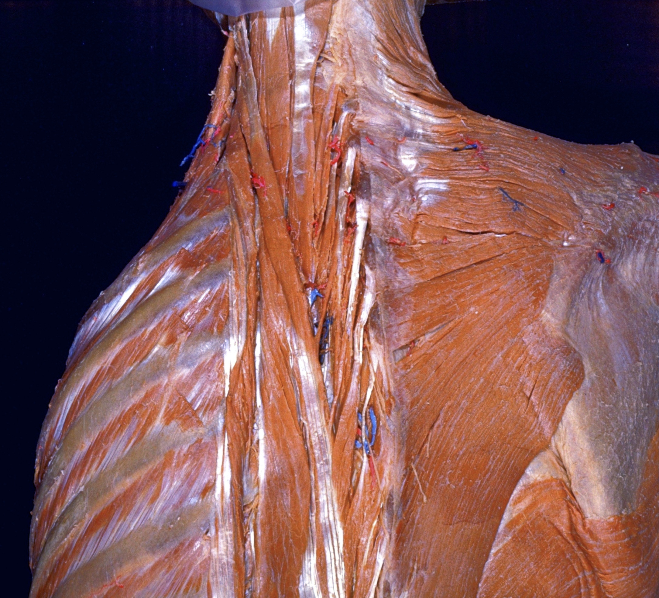
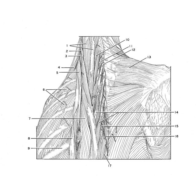
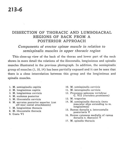
Dissection of thoracic and lumbosacral regions of back from a posterior approach
Components of erector spinae muscle in relation to semispinalis muscles in upper thoracic region
Stanford holds the copyright to the David L. Bassett anatomical images and has assigned
Creative Commons license Attribution-Share Alike 4.0 International to all of the images.
For additional information regarding use and permissions,
please contact Dr. Drew Bourn at dbourn@stanford.edu.
Image #213-6
 |  | ||||||||||||||||||||||||||||||||||||||
 |
|