Dissection of thoracic and lumbosacral regions of back from a posterior approach
Left serratus posterior inferior muscle
For permissions information regarding the use of these images,
please contact Drew Bourn at dbourn@stanford.edu
Image #213-1
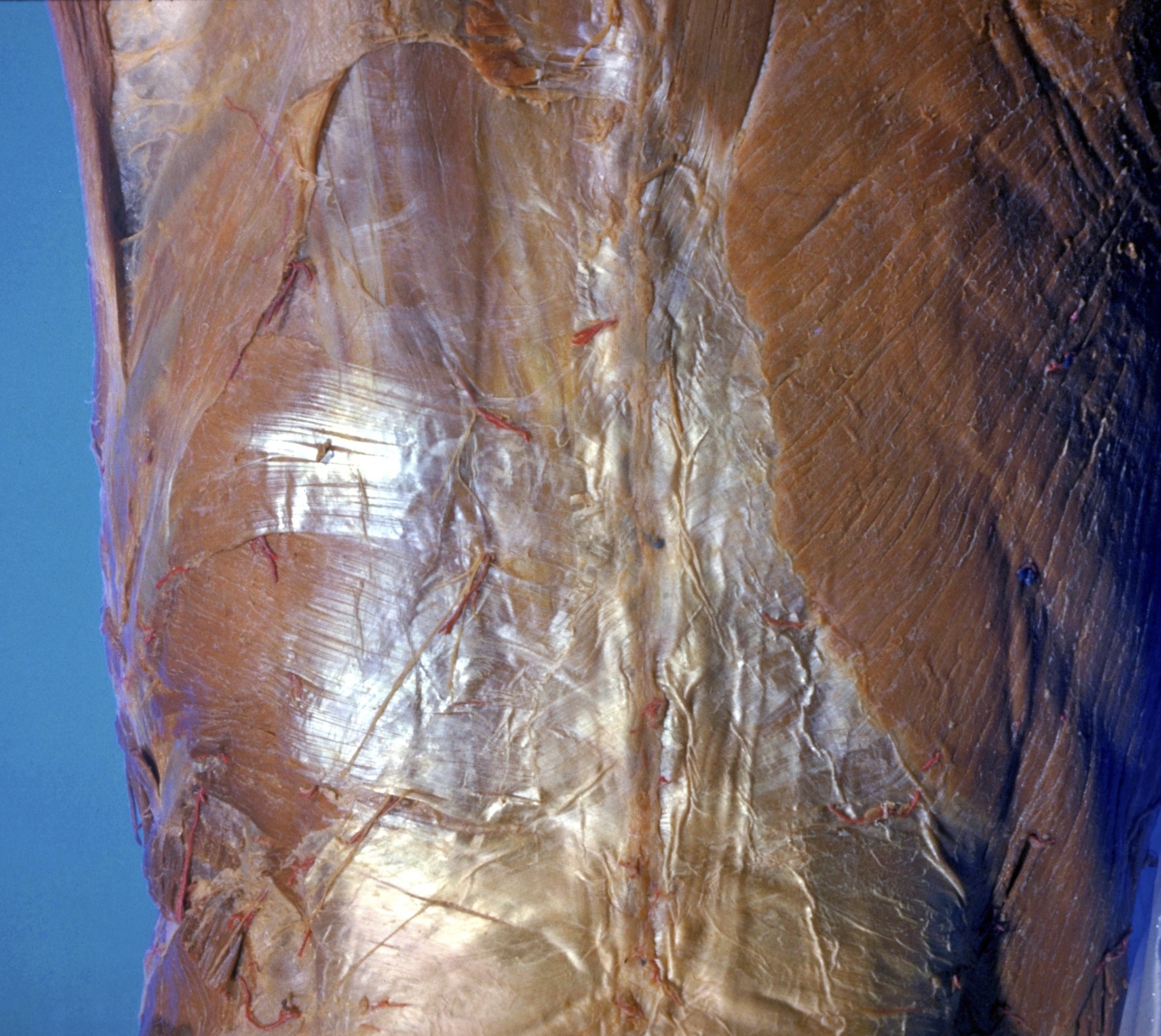
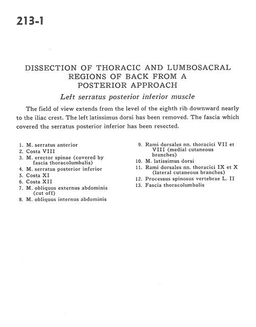
Dissection of thoracic and lumbosacral regions of back from a posterior approach
Left serratus posterior inferior muscle
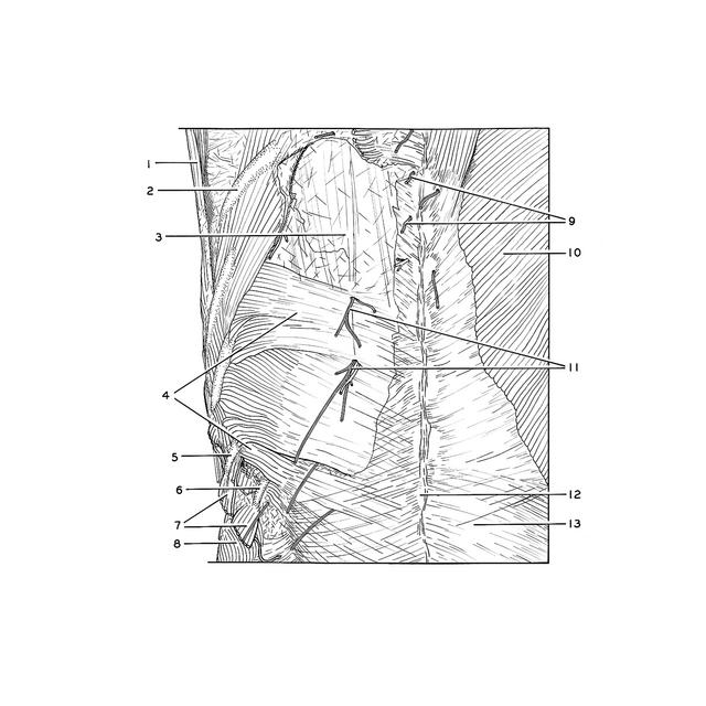
The field of view extends from the level of the eighth rib downward nearly to the iliac crest. The left latissimus dorsi has been removed. The fascia which covered the serratus posterior inferior has been resected.
1
.
Serratus anterior muscle
2
.
Rib VIII
3
.
Erector spinae muscle (covered by thoracolumbar fascia)
4
.
Serratus posterior inferior muscle
5
.
Rib XI
6
.
Rib XII
7
.
External oblique muscle (cut off)
8
.
Internal oblique muscle
9
.
Posterior branches of thoracic nerves VII and VIII (medial cutaneous branches)
10
.
Latissimus dorsi muscle
11
.
Posterior branches of thoracic nerves IX and X (lateral cutaneous branches)