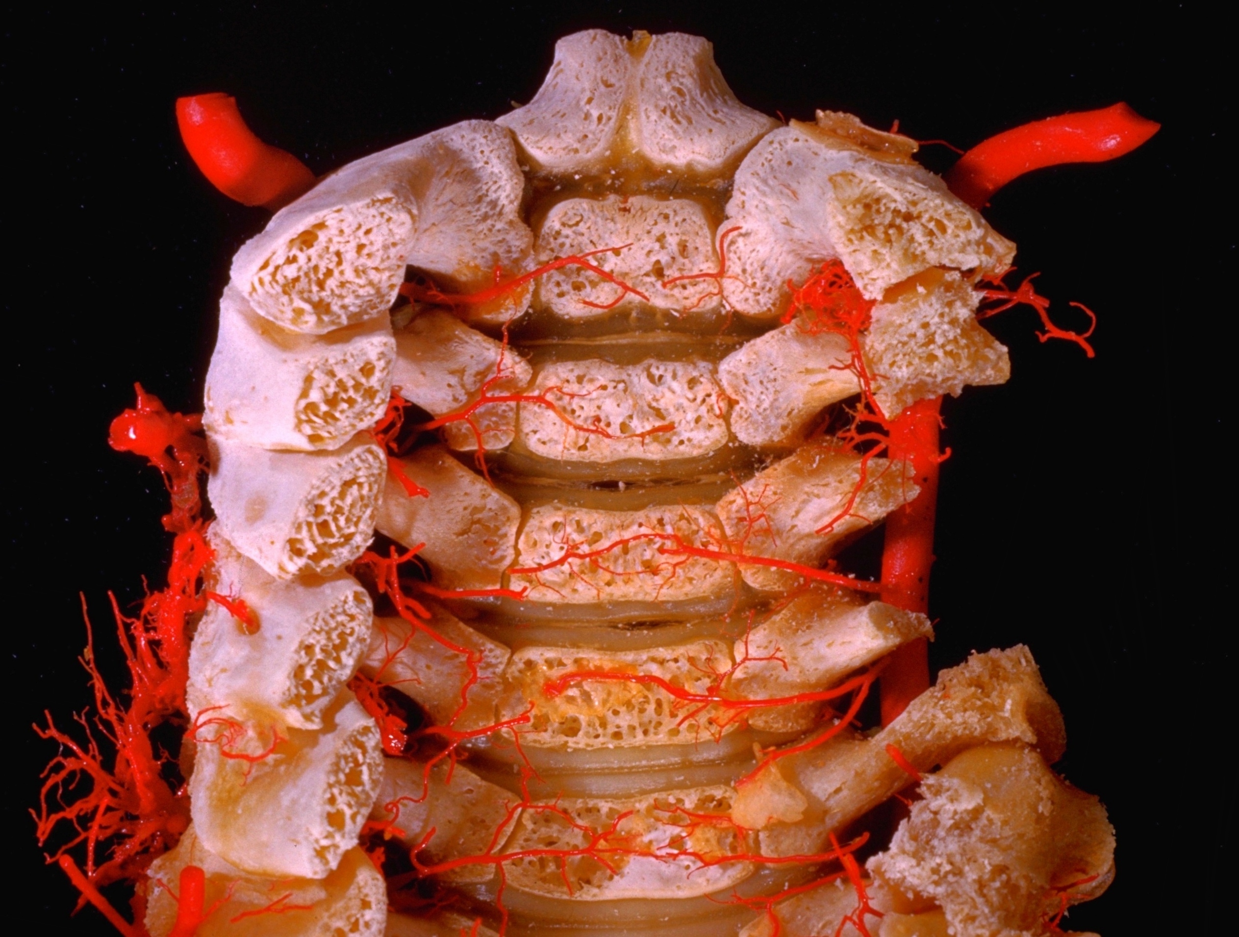
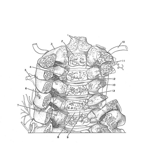
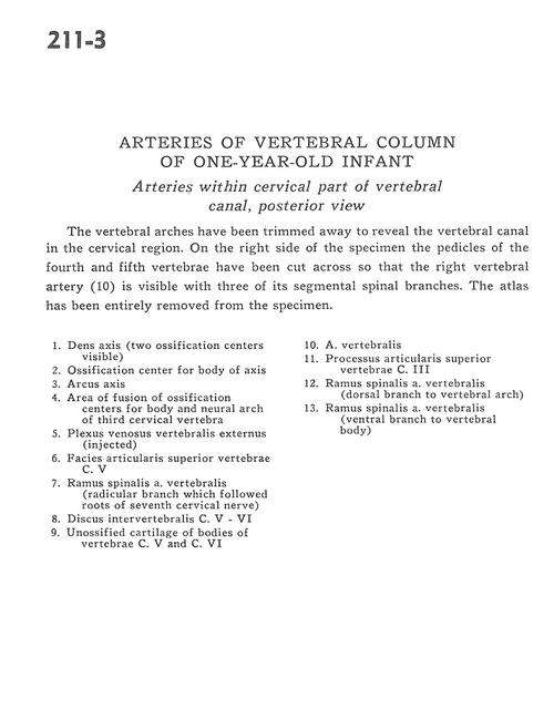
Arteries of vertebral column of one
Year-old infant - Arteries within cervical part of vertebral canal, posterior view
Stanford holds the copyright to the David L. Bassett anatomical images and has assigned
Creative Commons license Attribution-Share Alike 4.0 International to all of the images.
For additional information regarding use and permissions,
please contact Dr. Drew Bourn at dbourn@stanford.edu.
Image #211-3
 |  | ||||||||||||||||||||||||||||||
 |
|