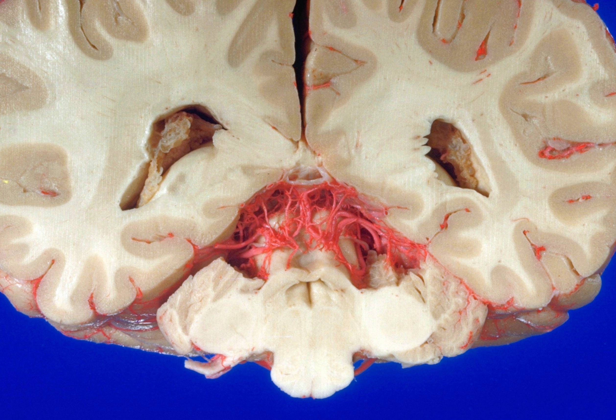
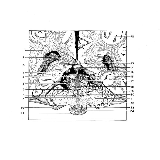
Exploration of a brain cut in frontal section at the splenium of the corpus callosum
Orientation view, cerebellum partly resected
Stanford holds the copyright to the David L. Bassett anatomical images and has assigned
Creative Commons license Attribution-Share Alike 4.0 International to all of the images.
For additional information regarding use and permissions,
please contact Dr. Drew Bourn at dbourn@stanford.edu.
Image #21-4
 |  | ||||||||||||||||||||||||||||||||||||||||||||||||||||
 |
|