Exploration of those parts of the brain supplied by the posterior cerebral artery
Lentiform nucleus and inferior occipitofrontal fasciculus
Stanford holds the copyright to the David L. Bassett anatomical images and has assigned
Creative Commons license Attribution-Share
Alike 4.0 International to all of the images.
For additional information regarding use and permissions,
please contact Dr. Drew Bourn at dbourn@stanford.edu.
Image #21-2
Exploration of those parts of the brain supplied by the posterior cerebral artery
Lentiform nucleus and inferior occipitofrontal fasciculus
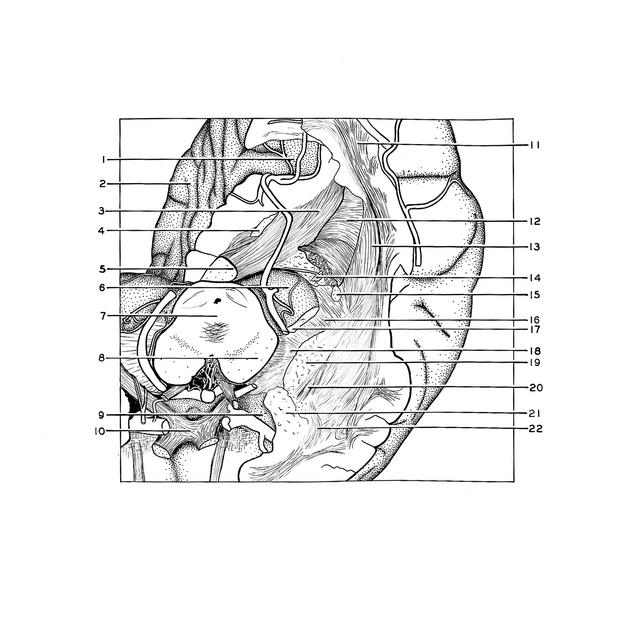
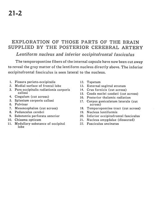
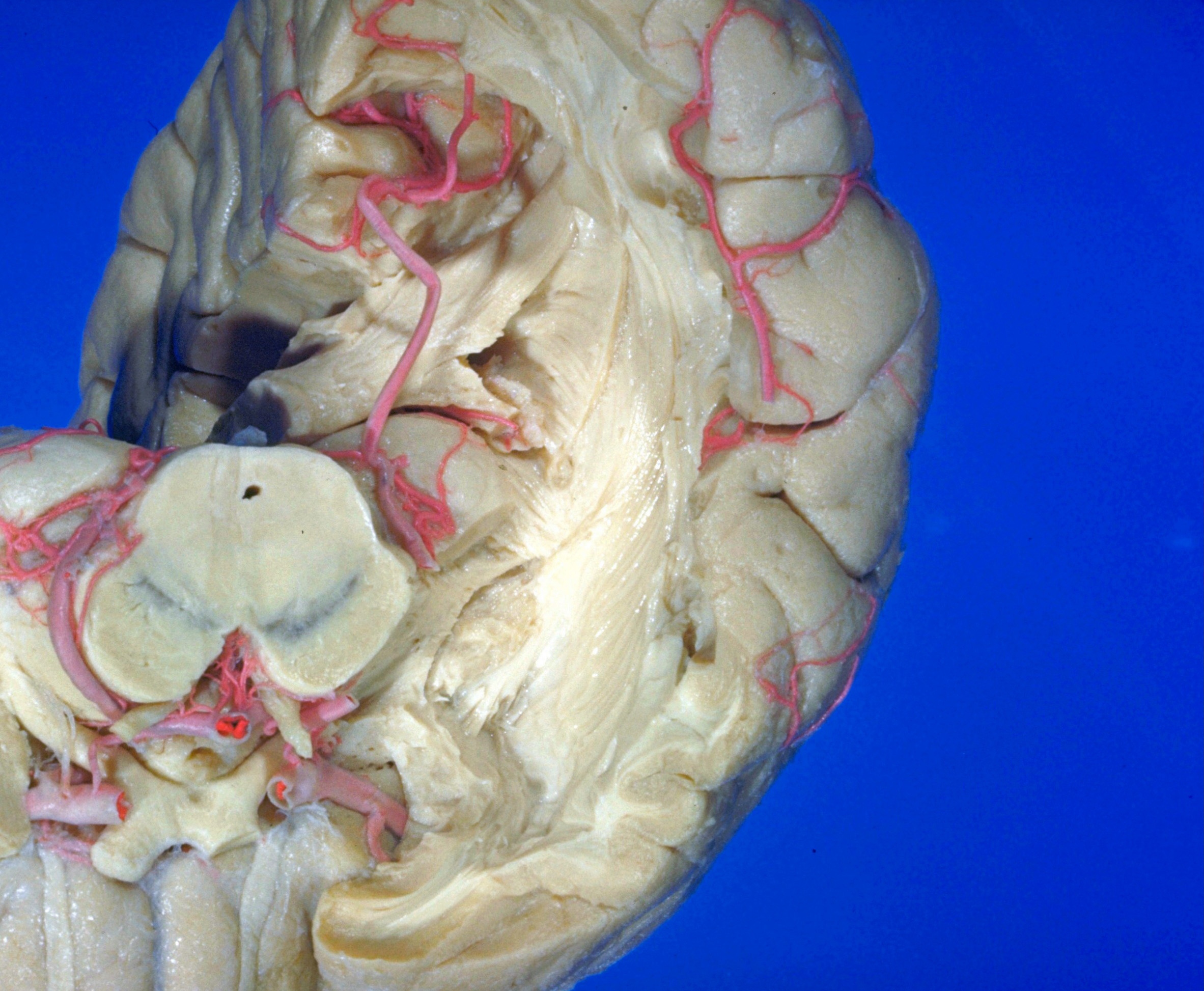
The temporopontine fibers of the internal capsule have now been cut away to reveal the gray matter of the lentiform nucleus directly above. The inferior occipitofrontal fasciculus is seen lateral to the nucleus.
- Parieto-occipital fissure
- Medial surface of frontal lobe
- Occipital part radiations of corpus callosum
- Cingulum (cut across)
- Corpus callosum (splenium)
- Pulvinar
- Mesencephalon (cut across)
- Cerebral peduncle
- Anterior perforated substance
- Optic chiasm
- Medullary substance of occipital lobe
- Tapetum
- External sagittal stratum
- Fornix (ems) (cut across)
- Caudate nucleus (tail) (cut across)
- Posterior thalamic radiation
- Lateral geniculate body (cut across)
- Temporopontine tract (cut across)
- Lentiform nucleus
- Inferior occipitofrontal fasciculus
- Amygdaloid nucleus (dissected)
- Uncinate fasciculus