X CLOSE

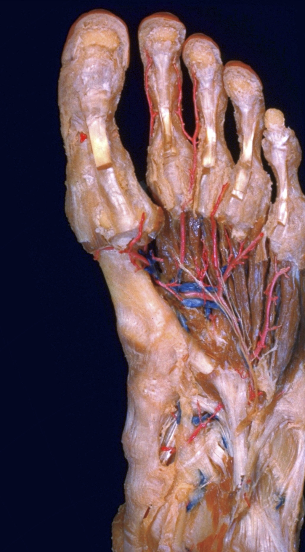
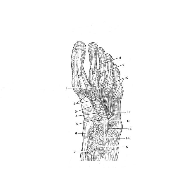
Dissection of plantar aspect of left foot

Interosseous muscles, general plantar view

For permissions information regarding the use of these images, please contact Drew Bourn at dbourn@stanford.edu

Image #202-2
Bassett ImageDiagram Image
Legend Image
Dissection of plantar aspect of left foot
Interosseous muscles, general plantar view
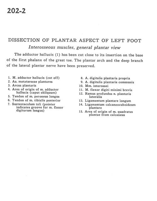
The adductor hallucis (1) has been cut close to its insertion on the base of the first phalanx of the great toe. The plantar arch and the deep branch of the lateral plantar nerve have been preserved.
1 . Adductor hallucis muscle (cut off)
2 . Metatarsal plantar arteries
3 . Plantar arch
4 . Area of origin of adductor hallucis muscle (oblique head)
5 . Tendon of peroneus longus muscle
6 . Tendon of tibialis posterior muscle
7 . Sustentaculum tali (pointer indicates groove for flexor digitorum longus muscle)
8 . Proper plantar digital artery
9 . Common plantar digital artery
10 . Interosseous muscle
11 . Flexor digiti minimi brevis muscle
12 . Deep branch lateral plantar nerve
13 . Long plantar ligament
14 . Calcaneocuboid plantar ligament
15 . Area of origin of quadratus plantae. muscle from calcaneus