X CLOSE

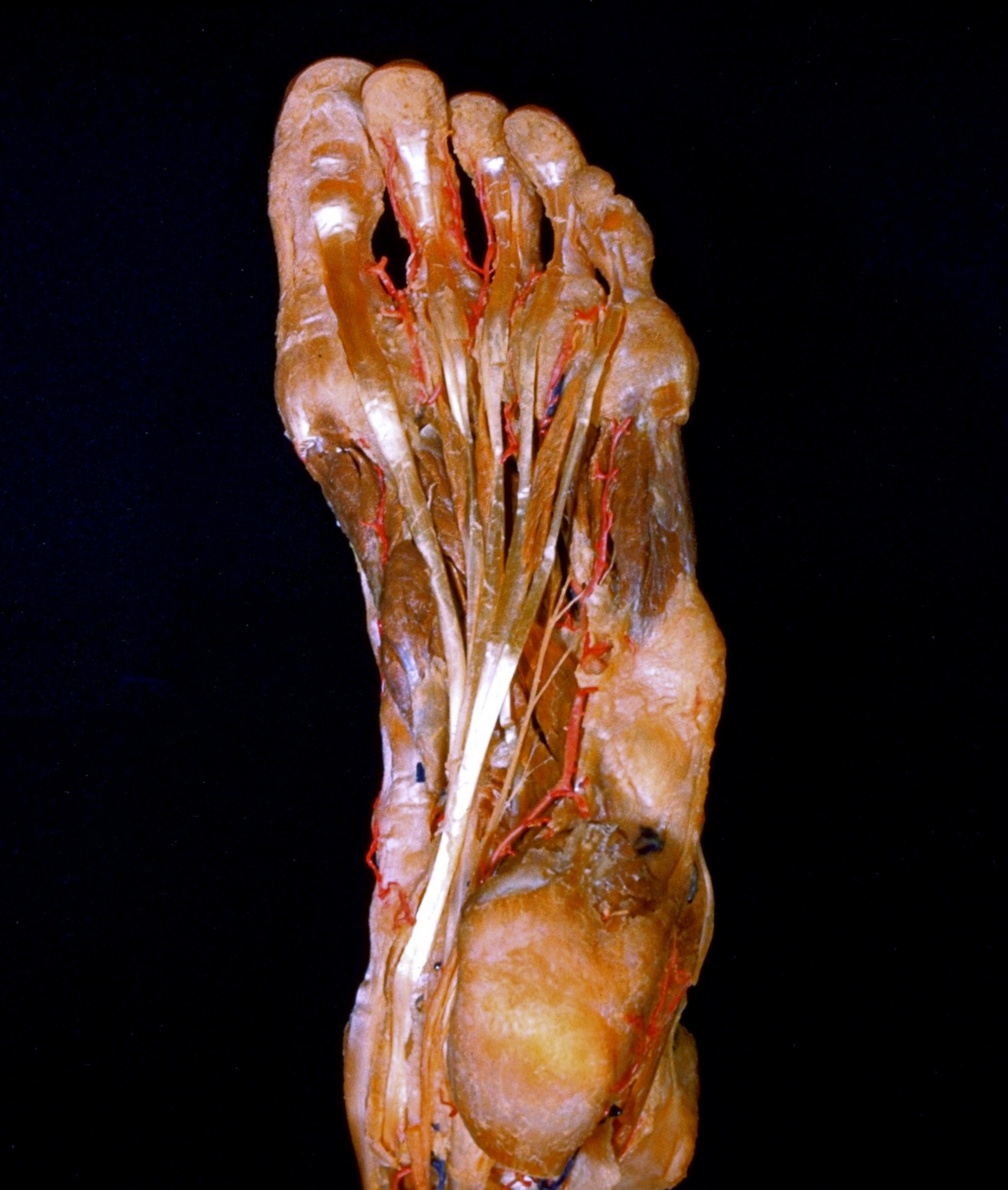
Dissection of plantar aspect of left foot

Long flexor tendons and lumbrical muscles

For permissions information regarding the use of these images, please contact Drew Bourn at dbourn@stanford.edu

Image #201-6
Bassett ImageDiagram Image
Legend Image
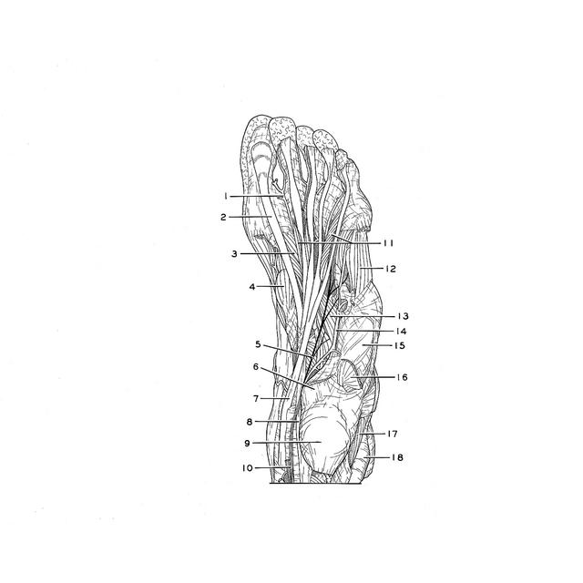
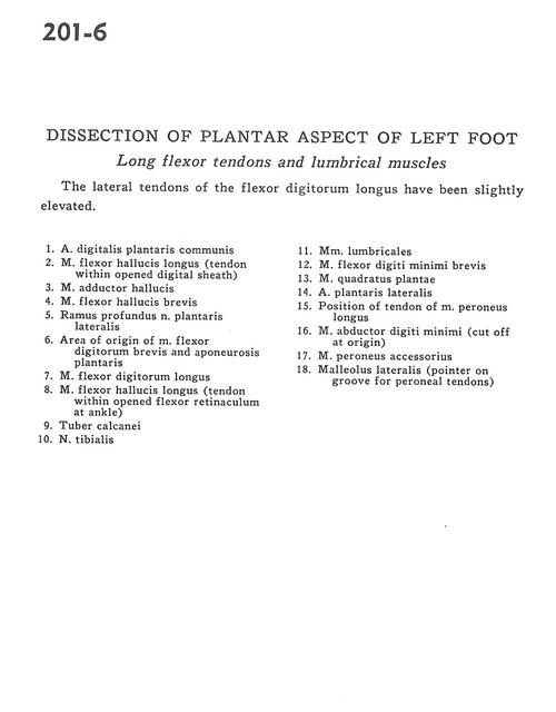
Dissection of plantar aspect of left foot
Long flexor tendons and lumbrical muscles
The lateral tendons of the flexor digitorum longus have been slightly elevated.
1 . Common plantar digital artery
2 . Flexor hallucis longus muscle (tendon within opened digital sheath)
3 . Adductor hallucis muscle
4 . Flexor hallucis brevis muscle
5 . Deep branch lateral plantar nerve
6 . Area of origin of flexor digitorum brevis muscle and plantar aponeurosis
7 . Flexor digitorum longus muscle
8 . Flexor hallucis longus muscle (tendon within opened flexor retinaculum at ankle)
9 . Tuberosity of calcaneus
10 . Tibial nerve
11 . Lumbrical muscles
12 . Flexor digiti minimi brevis muscle
13 . Quadratus plantae muscle
14 . Lateral plantar artery
15 . Position of tendon of peroneus longus muscle
16 . Abductor digiti minimi muscle (cut off at origin)
17 . Accessory peroneus muscle
18 . Lateral malleolus (pointer on groove for peroneal tendons)