X CLOSE

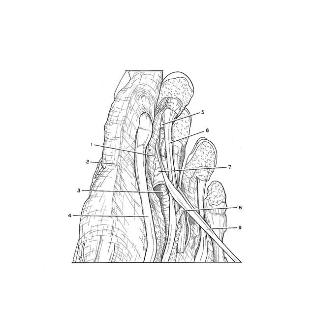
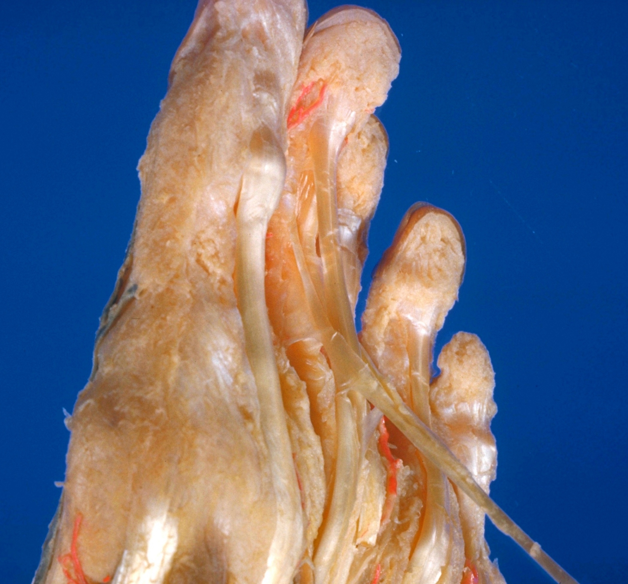
Dissection of plantar aspect of left foot

Flexor tendons of second toe elevated, medial close-up view

Stanford holds the copyright to the David L. Bassett anatomical images and has assigned Creative Commons license Attribution-Share Alike 4.0 International to all of the images.
For additional information regarding use and permissions, please contact Dr. Drew Bourn at dbourn@stanford.edu.

Image #201-2
Bassett ImageDiagram Image
Legend Image
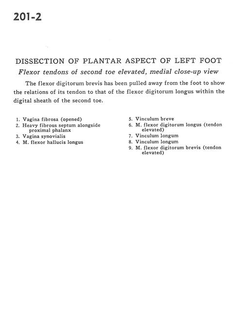
Dissection of plantar aspect of left foot
Flexor tendons of second toe elevated, medial close-up view
The flexor digitorum brevis has been pulled away from the foot to show the relations of its tendon to that of the flexor digitorum longus within the digital sheath of the second toe.
1 . Fibrous sheath (opened)
2 . Heavy fibrous septum alongside proximal phalanx
3 . Synovial sheath
4 . Flexor hallucis longus muscle
5 . Vinculum breve
6 . Flexor digitorum longus muscle (tendon elevated)
7 . Vinculum longum
8 . Vinculum longum
9 . Flexor digitorum brevis muscle (tendon elevated)