X CLOSE

Exploration of those parts of the brain supplied by the posterior cerebral artery

Looping fibers from stratum zonale of thalamus radiating into temporal and occipital lobes

Stanford holds the copyright to the David L. Bassett anatomical images and has assigned Creative Commons license Attribution-Share Alike 4.0 International to all of the images.
For additional information regarding use and permissions, please contact Dr. Drew Bourn at dbourn@stanford.edu.

Image #20-5
Bassett ImageDiagram Image
Legend Image
Exploration of those parts of the brain supplied by the posterior cerebral artery
Looping fibers from stratum zonale of thalamus radiating into temporal and occipital lobes
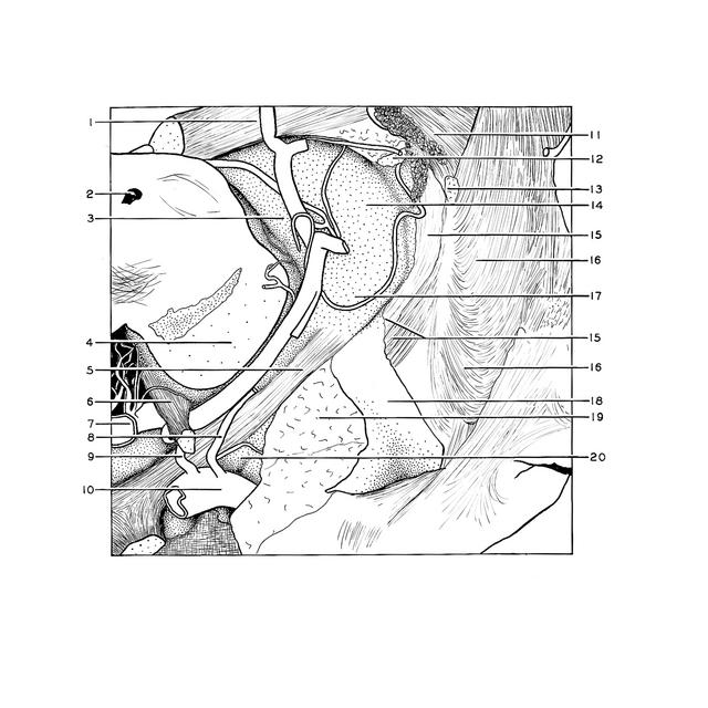
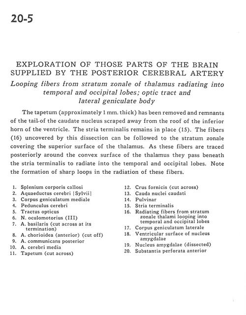
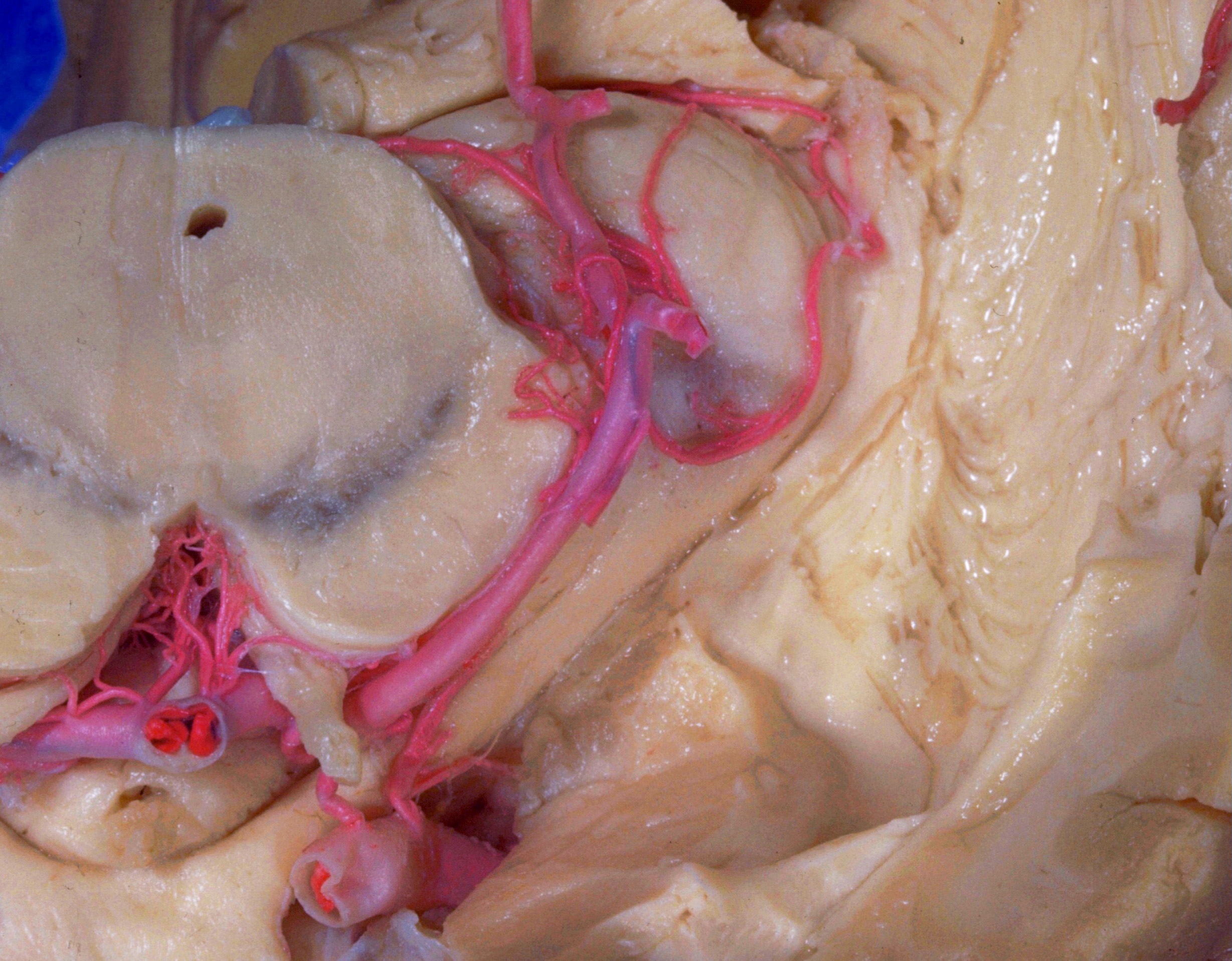
The tapetum (approximately 1 mm. thick) has been removed and remnants of the tail of the caudate nucleus scraped away from the roof of the inferior horn of the ventricle. The stria terminalis remains in place (15). The fibers (16) uncovered by this dissection can be followed to the stratum zonale covering the superior surface of the thalamus. As these fibers are traced posteriorly around the convex surface of the thalamus they pass beneath the stria terminalis to radiate into the temporal and occipital lobes. Note the formation of sharp loops in the radiation of these fibers.
1 . Corpus callosum (splenium)
2 . Cerebral aqueduct
3 . Medial geniculate body
4 . Cerebral peduncle
5 . Optic tract
6 . Oculomotor nerve (III)
7 . Basilar artery (cut across at its termination)
8 . Choroidal artery (anterior) (cut off)
9 . Posterior communicating artery
10 . Middle cerebral artery
11 . Tapetum (cut across)
12 . Fornix (crus) (cut across)
13 . Caudate nucleus (tail)
14 . Pulvinar
15 . Stria terminalis
16 . Radiating fibers from stratum zonale thalami looping into temporal and occipital lobes
17 . Lateral geniculate body
18 . Ventricular surface of amygdala
19 . Amygdaloid nucleus (dissected)
20 . Anterior perforated substance