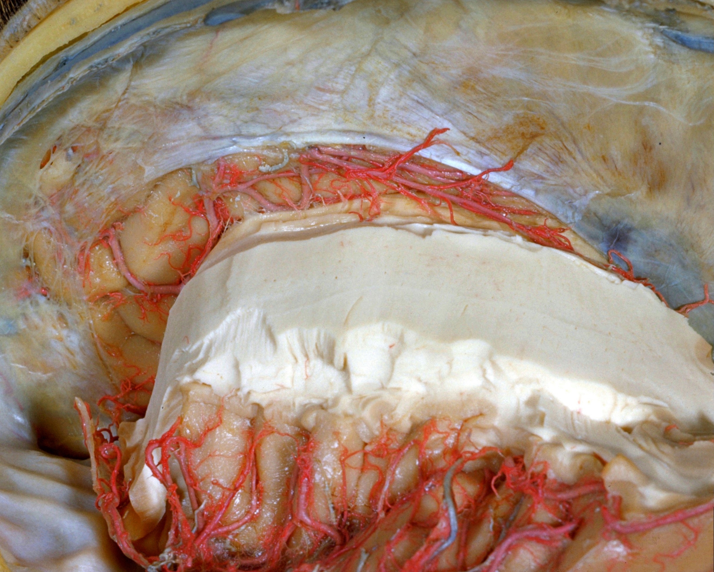
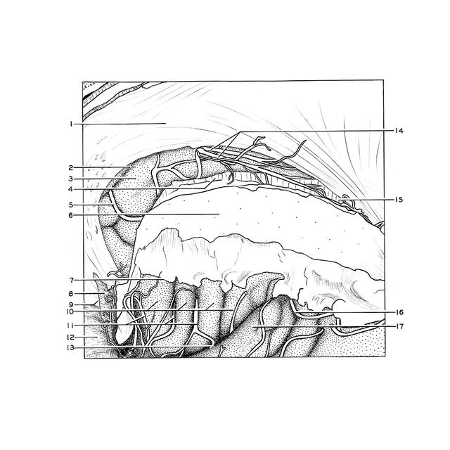
Exploration of the meninges and brain in situ
Pericallosal arteries (both arising from right anterior cerebral artery)
Stanford holds the copyright to the David L. Bassett anatomical images and has assigned
Creative Commons license Attribution-Share Alike 4.0 International to all of the images.
For additional information regarding use and permissions,
please contact Dr. Drew Bourn at dbourn@stanford.edu.
Image #2-2
 |  | ||||||||||||||||||||||||||||||||||||||
 |
|