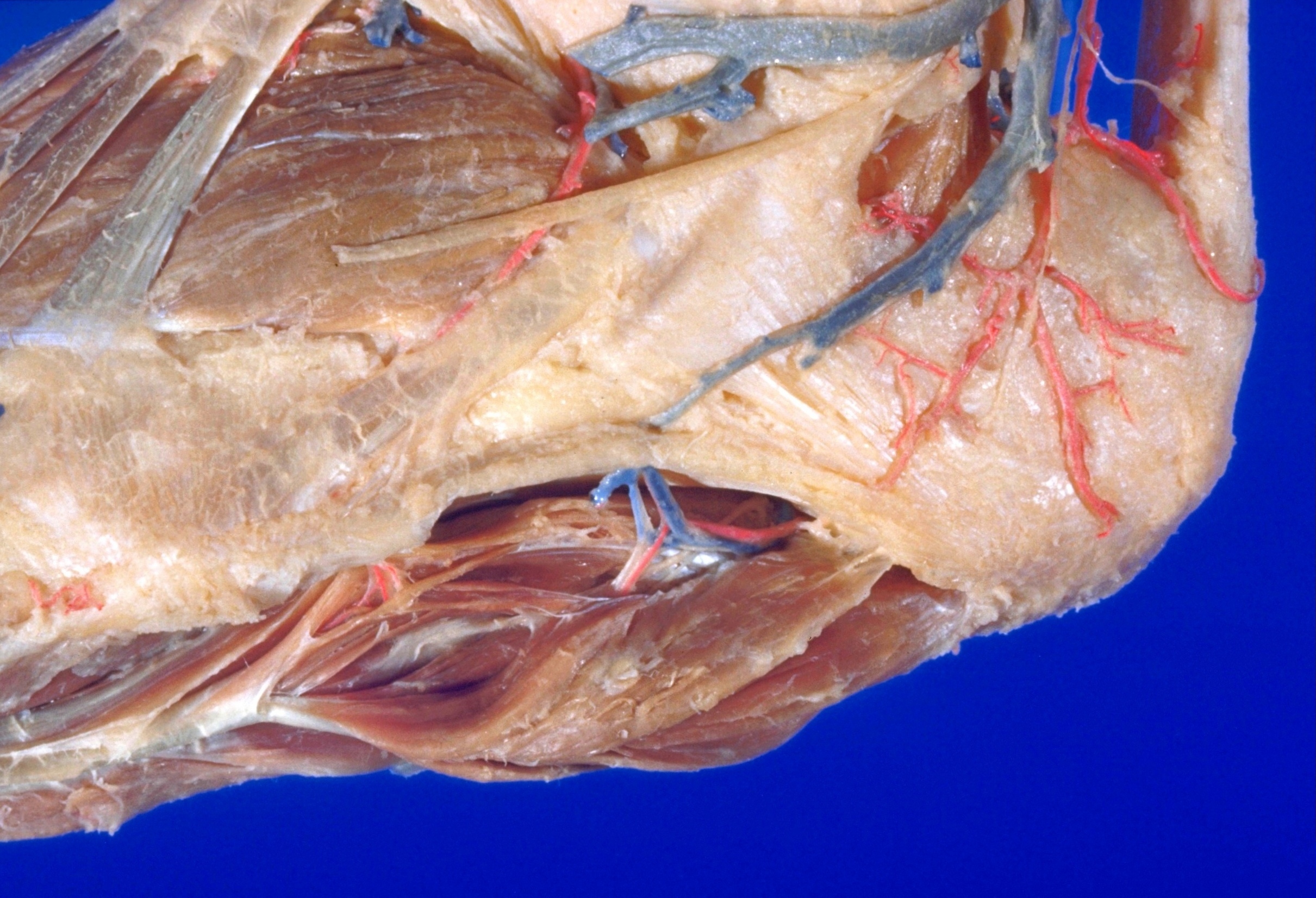
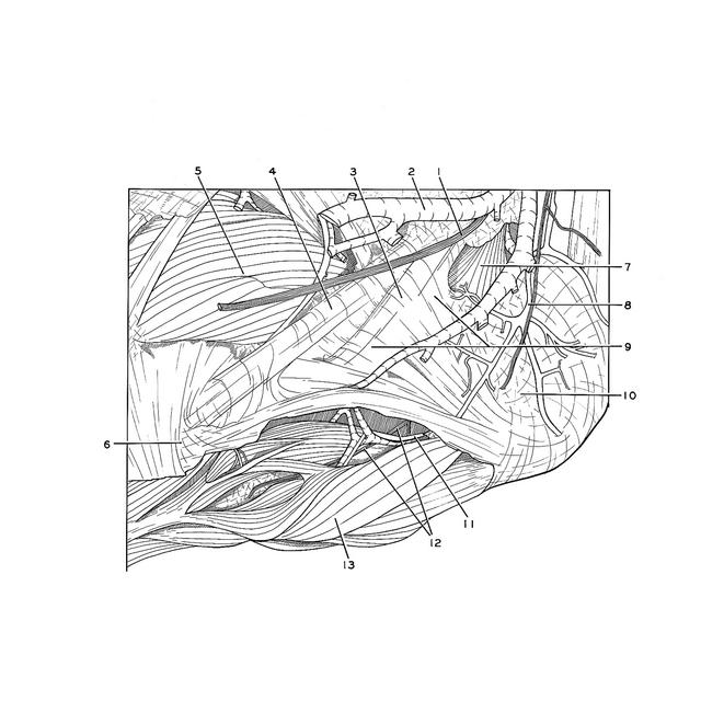
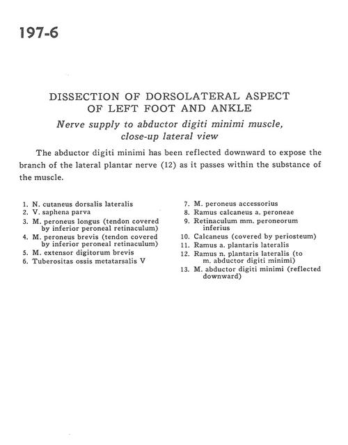
Dissection of dorsolateral aspect of left foot and ankle
Nerve supply to abductor digiti minimi muscle, close-up lateral view
Stanford holds the copyright to the David L. Bassett anatomical images and has assigned
Creative Commons license Attribution-Share Alike 4.0 International to all of the images.
For additional information regarding use and permissions,
please contact Dr. Drew Bourn at dbourn@stanford.edu.
Image #197-6
 |  | ||||||||||||||||||||||||||||||
 |
|