X CLOSE

Dissection of posterior aspect of left leg

Nerve supply to soleus muscle

Stanford holds the copyright to the David L. Bassett anatomical images and has assigned Creative Commons license Attribution-Share Alike 4.0 International to all of the images.
For additional information regarding use and permissions, please contact Dr. Drew Bourn at dbourn@stanford.edu.

Image #195-2
Bassett ImageDiagram Image
Legend Image
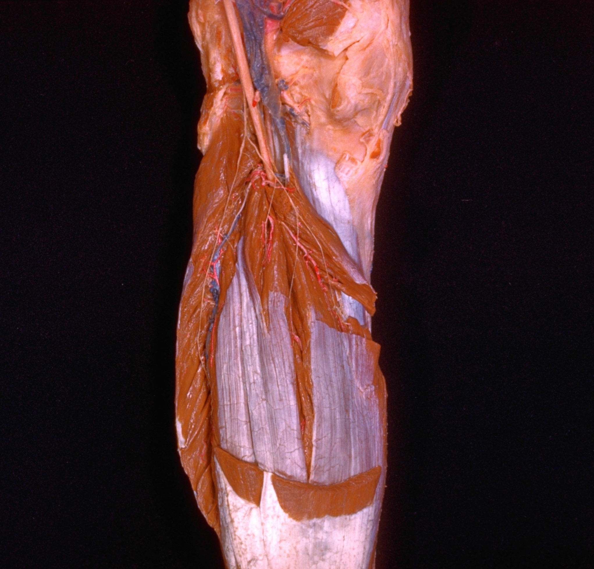
Dissection of posterior aspect of left leg
Nerve supply to soleus muscle
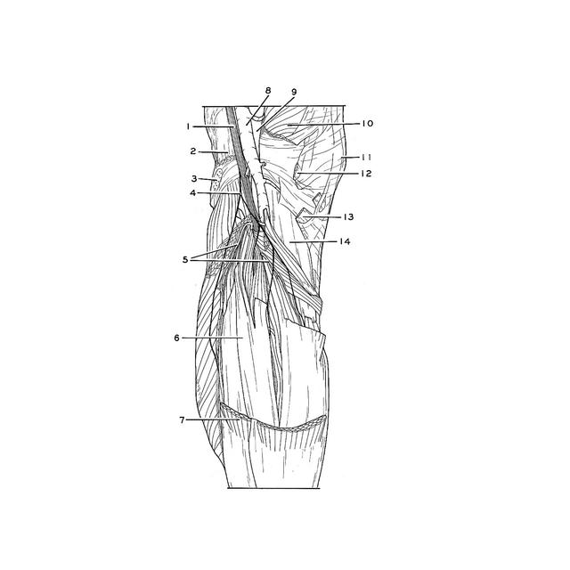
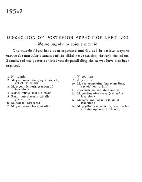
The muscle fibres have been separated and divided in various ways to expose the muscular branches of the tibial nerve passing through the soleus. Branches of the posterior tibial vessels paralleling the nerves have also been exposed.
1 . Tibial nerve
2 . Gastrocnemius muscle (lateral head cut off at origin)
3 . Biceps femoris muscle (tendon of insertion)
4 . Muscular branch of tibial nerve
5 . Muscular branch posterior tibial artery
6 . Soleus muscle (dissected)
7 . Gastrocnemius muscle (cut off)
8 . Popliteal vein
9 . Popliteal artery
10 . Gastrocnemius muscle (caput mediale, cut off near origin)
11 . Medial epicondyle of femur
12 . Semimembranosus muscle (cut off at insertion)
13 . Semitendinosus muscle (cut off at insertion)
14 . Popliteus muscle (covered by vertically directed aponeurotic fibers)