X CLOSE

Dissection of anterior aspect of left leg

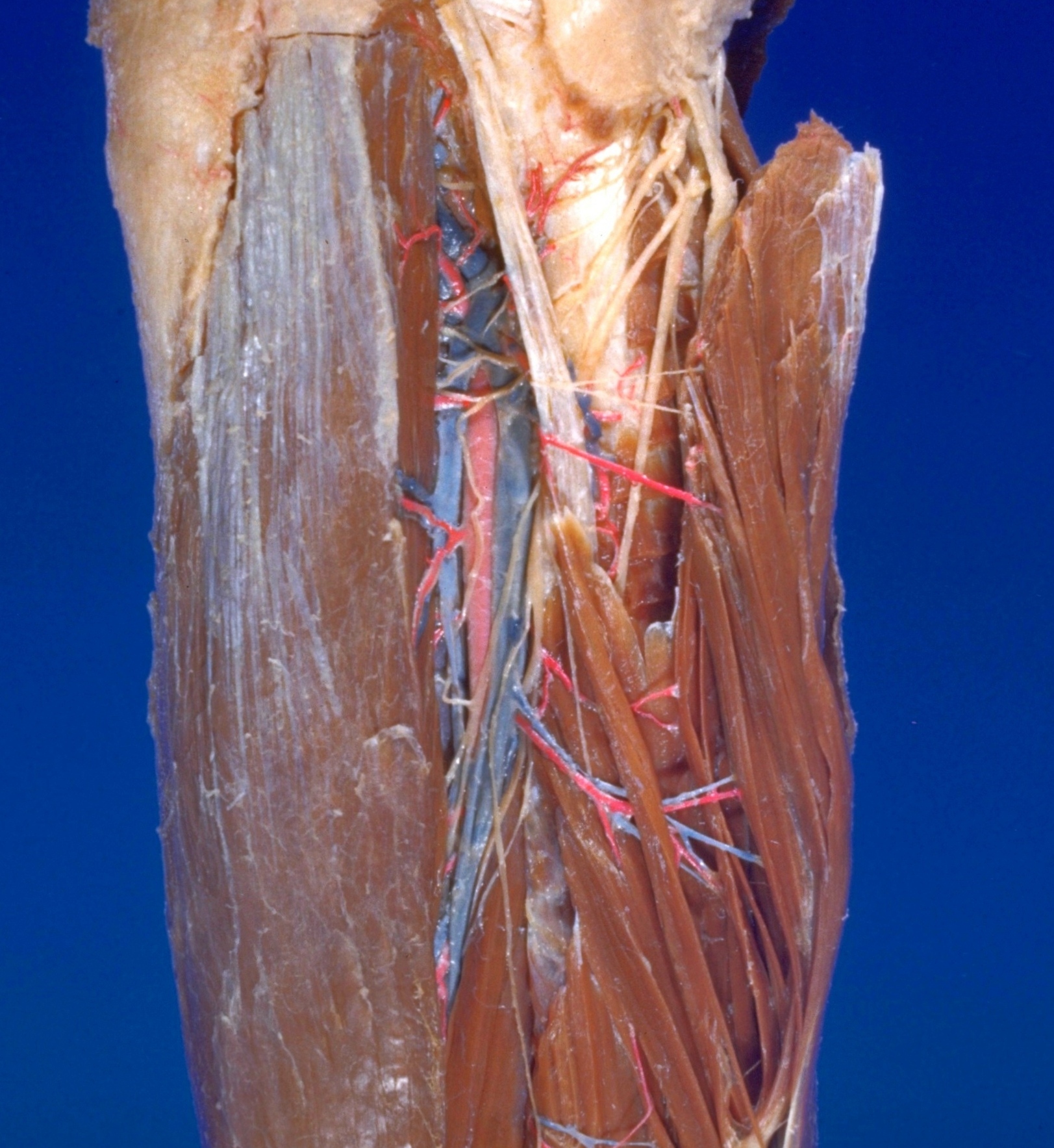
Nerve supply to extensor digitorum longus, close-up view of upper part of leg

For permissions information regarding the use of these images, please contact Drew Bourn at dbourn@stanford.edu

Image #192-3
Bassett ImageDiagram Image
Legend Image
Dissection of anterior aspect of left leg
Nerve supply to extensor digitorum longus, close-up view of upper part of leg
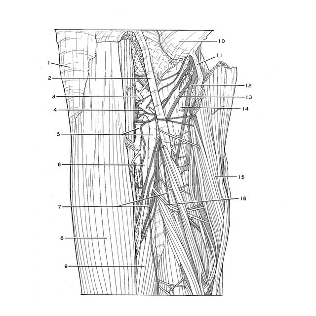
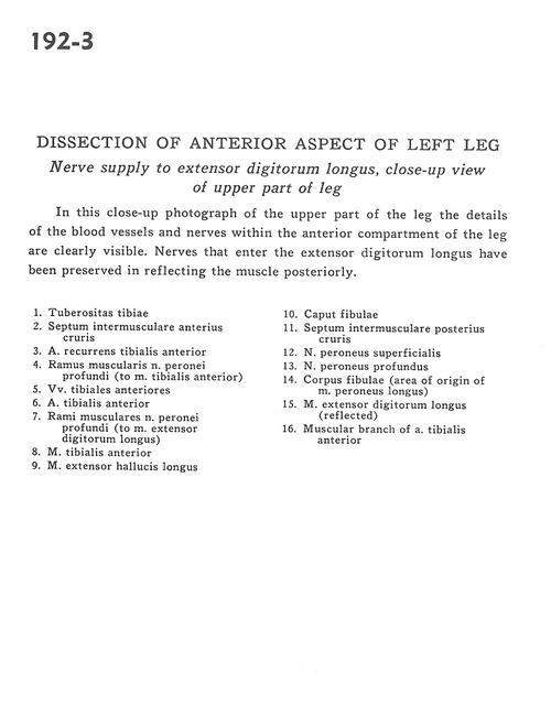
This close-up photograph of the upper part of the leg the details of the blood vessels and nerves within the anterior compartment of the leg are clearly visible. Nerves that enter the extensor digitorum longus have been preserved in reflecting the muscle posteriorly.
1 . Tibial tuberosity
2 . Anterior intermuscular septum
3 . Anterior tibial recurrent artery
4 . Muscular branch of deep peroneal nerve (to tibialis anterior muscle)
5 . Anterior tibial veins
6 . Anterior tibial artery
7 . Muscular branch of deep peroneal nerve (to extensor digitorum longus muscle)
8 . Tibialis anterior muscle
9 . Extensor hallucis longus muscle
10 . Head of fibula
11 . Posterior intermuscular septum
12 . Superficial peroneal nerve
13 . Deep peroneal nerve
14 . Body of fibula (area of origin of peroneus longus muscle)
15 . Extensor digitorum longus muscle (reflected)
16 . Muscular branch of anterior tibial artery