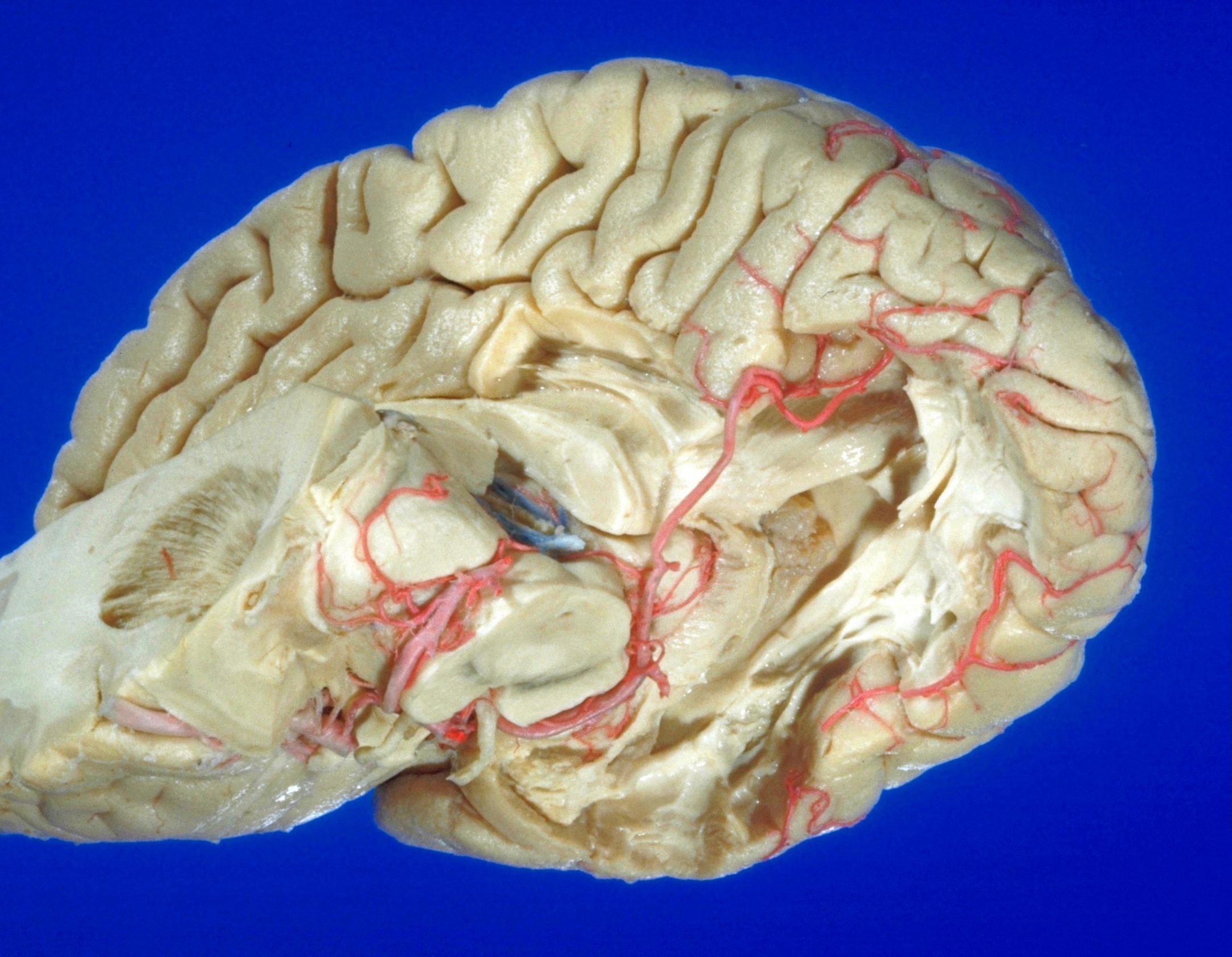
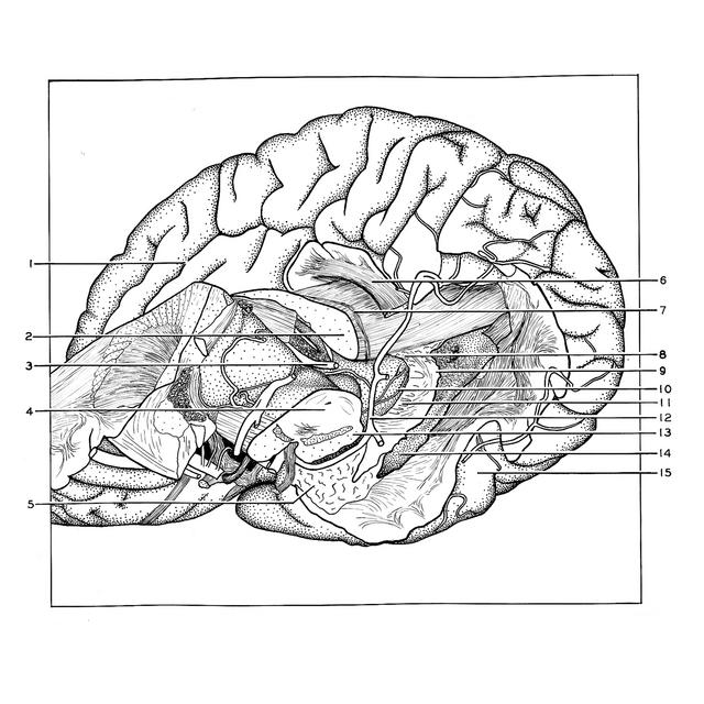
Exploration of those parts of the brain supplied by the posterior cerebral artery
General view of relations of dentate fascia and hippocampus
Stanford holds the copyright to the David L. Bassett anatomical images and has assigned
Creative Commons license Attribution-Share Alike 4.0 International to all of the images.
For additional information regarding use and permissions,
please contact Dr. Drew Bourn at dbourn@stanford.edu.
Image #19-7
 |  | ||||||||||||||||||||||||||||||||||
 |
|