X CLOSE

Dissection of knee

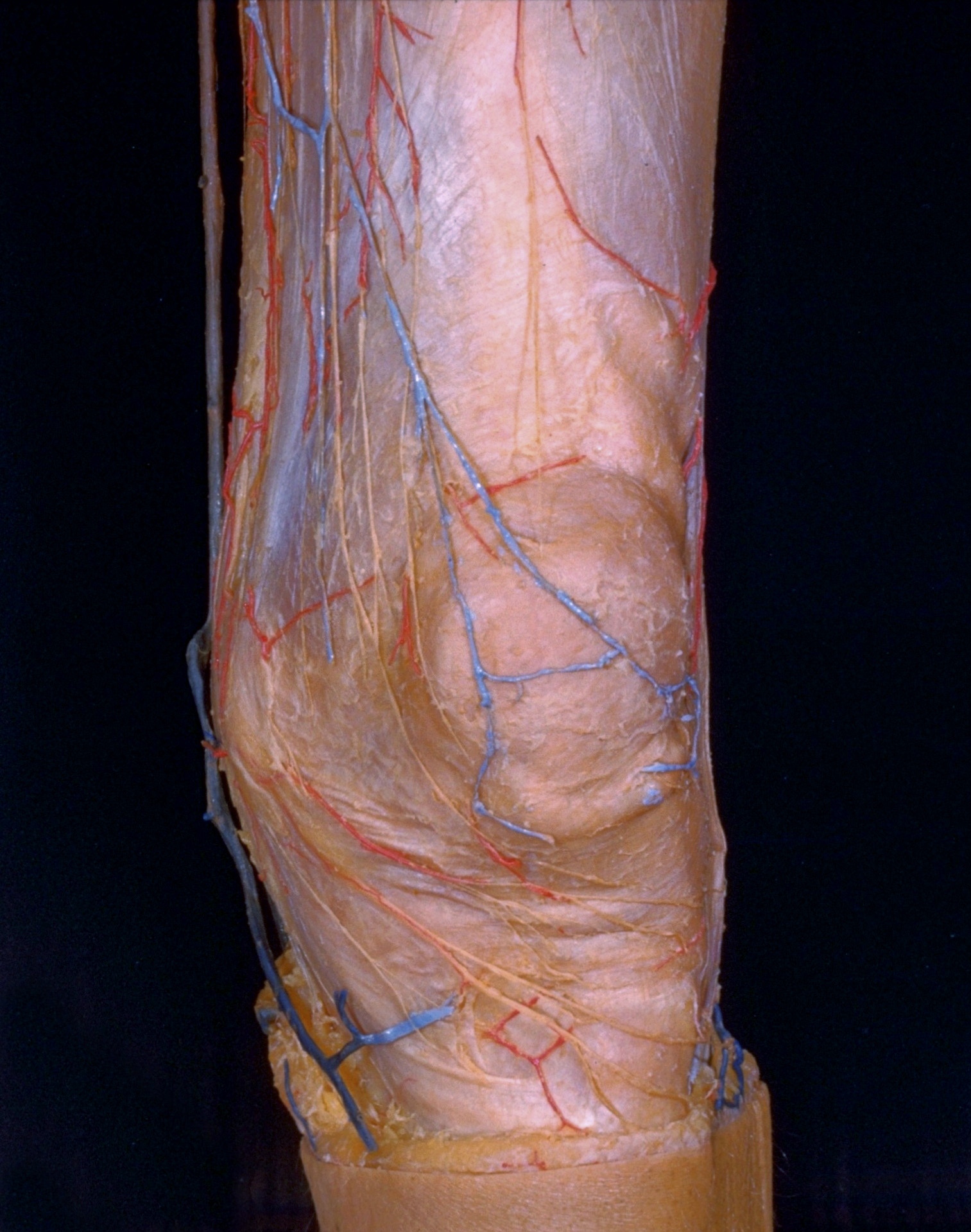
Superficial vessels and nerves of left knee, anterior view

For permissions information regarding the use of these images, please contact Drew Bourn at dbourn@stanford.edu

Image #189-4
Bassett ImageDiagram Image
Legend Image
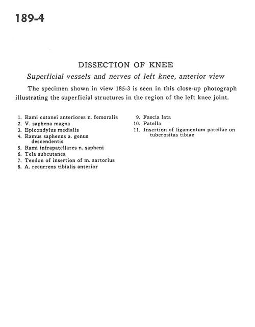
Dissection of knee
Superficial vessels and nerves of left knee, anterior view
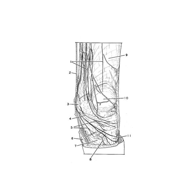
The specimen shown in view 185-3 is seen in this close-up photograph illustrating the superficial structures in the region of the left knee joint.
1 . Cutaneous branches of anterior femoral nerve
2 . Greater saphenous vein
3 . Medial epicondyle
4 . Saphenous branch of descending genicular artery
5 . Infrapatellar branch of saphenous nerve
6 . Tela subcutanea
7 . Tendon of insertion of sartorius muscle
8 . Anterior tibial recurrent artery
9 . Fascia lata
10 . Patella
11 . Insertion of patellar ligament on tibial tuberosity