For permissions information regarding the use of these images,
please contact Drew Bourn at dbourn@stanford.edu
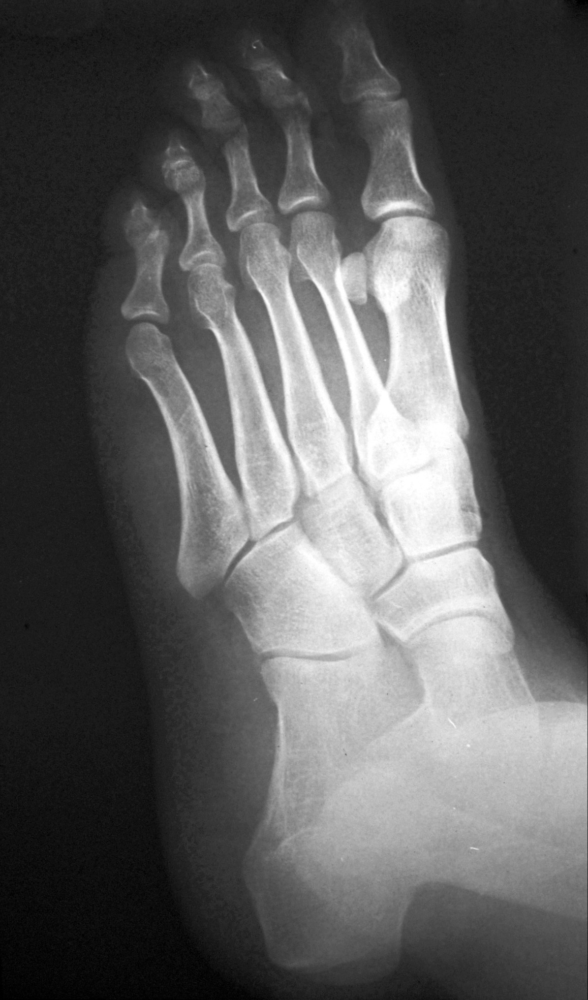
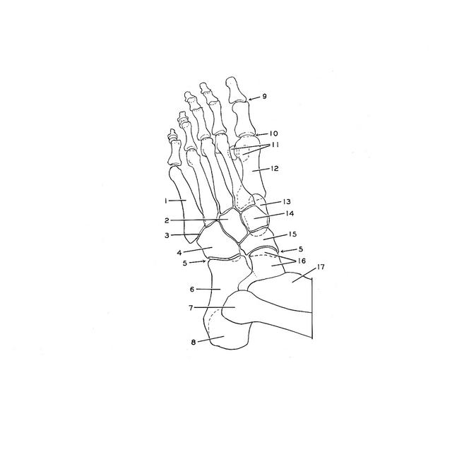
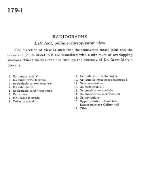
Image #179-1
Radiography
Left foot, oblique dorsoplantar view
The direction of view is such that the transverse tarsal joint and the bones and joints distal to it are visualized with a minimum of overlapping shadows. This film was obtained through the courtesy of Dr. Grant Melvin Stevens.
1
.
5th metatarsal bone
2
.
Lateral cuneiform bone
3
.
Tarsometatarsal articulation
4
.
Cuboid bone
5
.
Transverse tarsal articulation
6
.
Calcaneus
7
.
Lateral malleolus
8
.
Tuberosity of calcaneus
9
.
Interphalangeal articulation
10
.
1st metatarsophalangeal articulations
11
.
Sesamoid bone
12
.
Metatarsal bone
13
.
Medial cuneiform bone
14
.
Intermediate cuneiform bone
15
.
Navicular bone
16
.
Upper pointer: Head of talus Lower pointer: Neck of talus