X CLOSE

Osteology

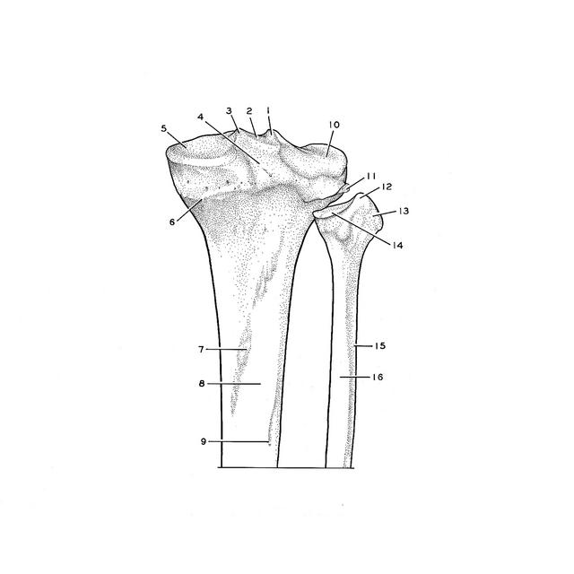
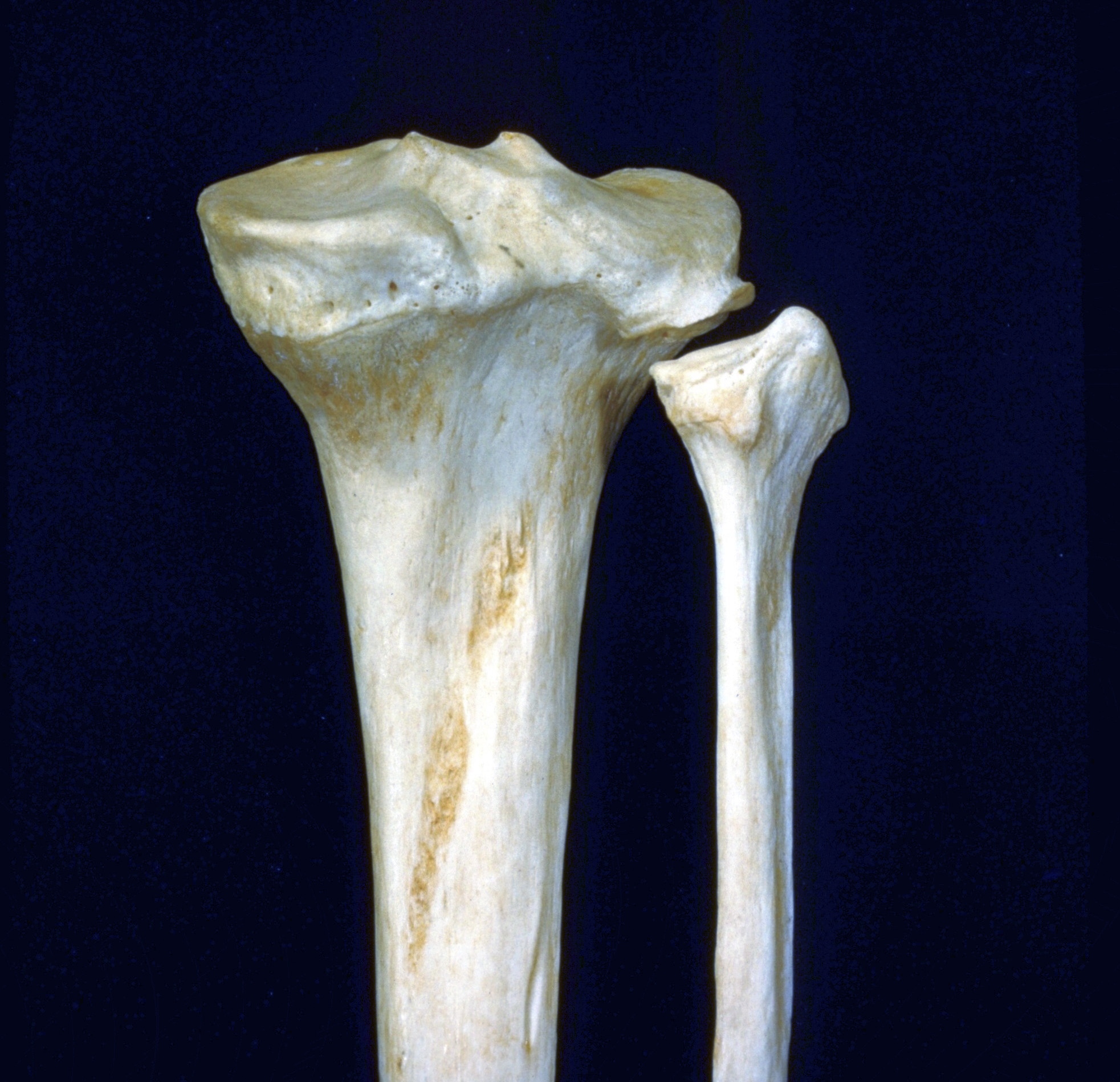
Right tibia and fibula, posterior view of upper parts

Stanford holds the copyright to the David L. Bassett anatomical images and has assigned Creative Commons license Attribution-Share Alike 4.0 International to all of the images.
For additional information regarding use and permissions, please contact Dr. Drew Bourn at dbourn@stanford.edu.

Image #176-7
Bassett ImageDiagram Image
Legend Image
Osteology
Right tibia and fibula, posterior view of upper parts
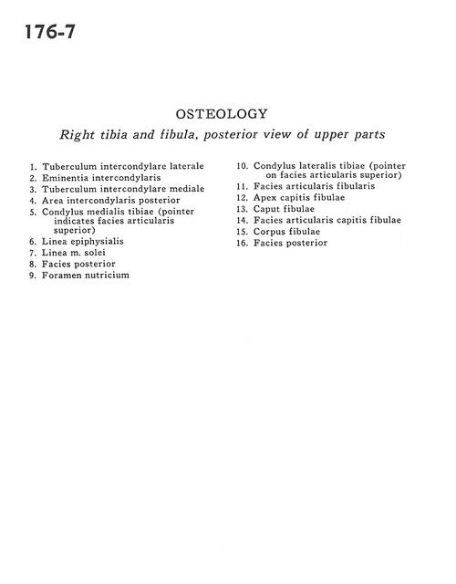
1 . Lateral intercondylar tubercle
2 . Intercondylar eminence
3 . Medial intercondylar tubercle
4 . Posterior intercondylar area
5 . Medial condyle of tibia (pointer indicates superior articular surface)
6 . Epiphysial line
7 . Soleal line
8 . Posterior surface
9 . Nutrient foramen
10 . Lateral condyle of tibia (pointer on superior articular surface)
11 . Articular surface of fibula
12 . Apex of fibula
13 . Head of fibula
14 . Articular surface of head of fibula
15 . Body of fibula
16 . Posterior surface