X CLOSE

Osteology

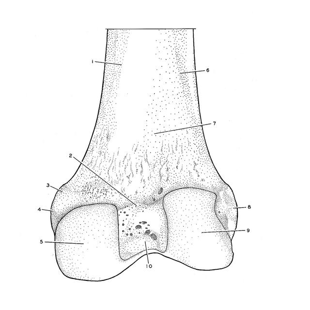
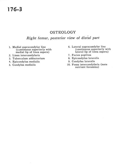
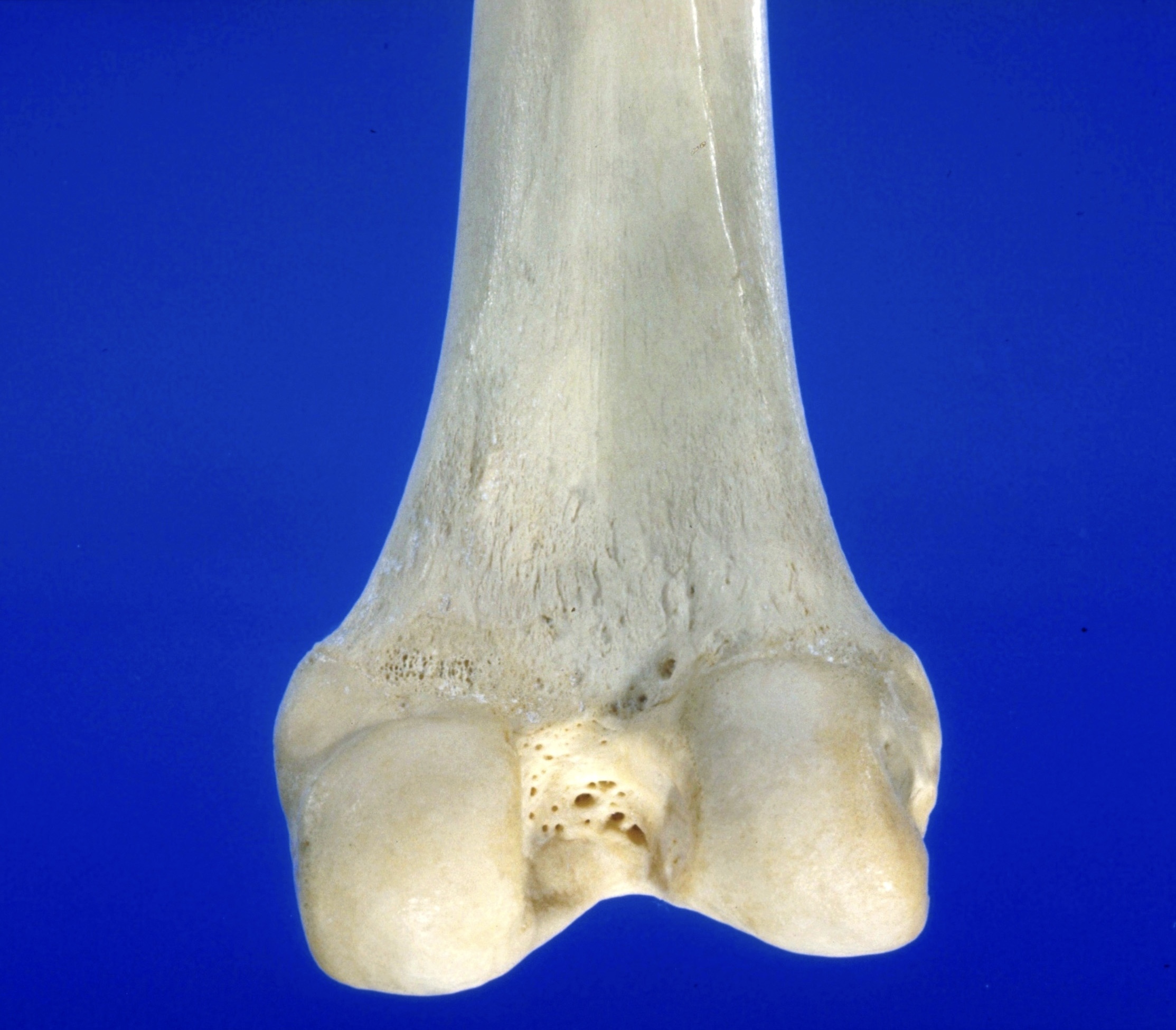
Right femur, posterior view of distal part

Stanford holds the copyright to the David L. Bassett anatomical images and has assigned Creative Commons license Attribution-Share Alike 4.0 International to all of the images.
For additional information regarding use and permissions, please contact Dr. Drew Bourn at dbourn@stanford.edu.

Image #176-3
Bassett Image
Diagram Image
Legend Image
Osteology
Right femur, posterior view of distal part
  1. Medial supracondylar line (continuous superiorly with medial lip of linea aspera)
  2. Intercondylar line
  3. Adductor tubercle
  4. Medial epicondyle
  5. Medial condyle
  6. Lateral supracondylar line (continuous superiorly with lateral lip of linea aspera)
  7. Popliteal surface
  8. Lateral epicondyle
  9. Lateral condyle
  10. Intercondylar fossa (note nutrient foramen)