Osteology
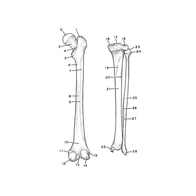
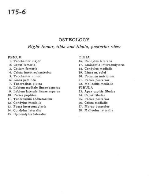
Right femur, tibia and fibula, posterior view
Stanford holds the copyright to the David L. Bassett anatomical images and has assigned
Creative Commons license Attribution-Share
Alike 4.0 International to all of the images.
For additional information regarding use and permissions,
please contact Dr. Drew Bourn at dbourn@stanford.edu.
Image #175-6
Osteology
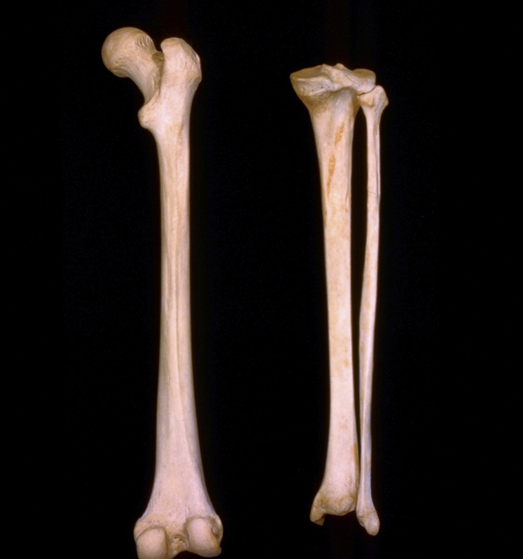
Right femur, tibia and fibula, posterior view
- Femur
- Greater trochanter
- Head of femur
- Neck of femur
- Intertrochanteric crest
- Lesser trochanter
- Pectineal line
- Gluteal tuberosity
- Medial lip of linea aspera
- Lateral lip of linea aspera
- Popliteal surface
- Adductor tubercle
- Medial condyle
- Intercondylar fossa
- Lateral condyle
- Lateral epicondyle
- Tibia
- Lateral condyle
- Intercondylar eminence
- Medial condyle
- Soleal line
- Nutrient foramen
- Posterior surface
- Medial malleolus
- Fibula
- Apex of fibula
- Head of fibula
- Posterior surface
- Medial crest
- Posterior border
- Lateral malleolus