X CLOSE

Osteology

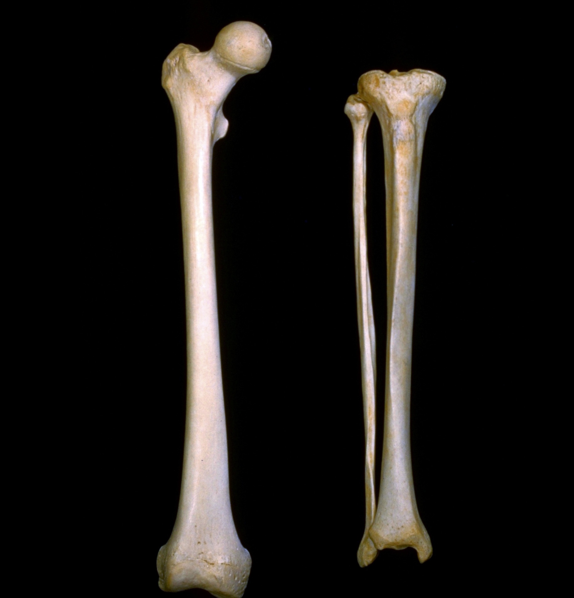
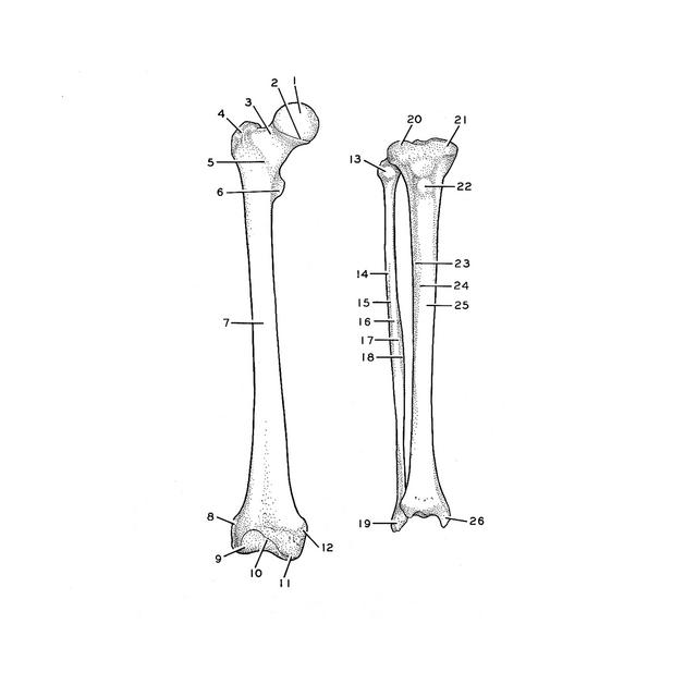
Right femur, tibia and fibula, anterior view

For permissions information regarding the use of these images, please contact Drew Bourn at dbourn@stanford.edu

Image #175-5
Bassett ImageDiagram Image
Legend Image
Osteology
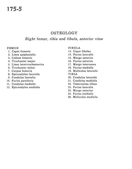
Right femur, tibia and fibula, anterior view
- . Femur
1 . Head of femur
2 . Epiphysial line
3 . Neck of femur
4 . Greater trochanter
5 . Intertrochanteric line
6 . Lesser trochanter
7 . Body of femur
8 . Lateral epicondyle
9 . Lateral condyle
10 . Patellar surface
11 . Medial condyle
12 . Medial epicondyle
- . Fibula
13 . Head of fibula
14 . Lateral surface
15 . Anterior border
16 . Anterior surface
17 . Interosseous border
18 . Medial surface
19 . Lateral malleolus
- . Tibia
20 . Lateral condyle
21 . Medial condyle
22 . Tibial tuberosity
23 . Lateral surface
24 . Anterior border
25 . Medial surface
26 . Medial malleolus