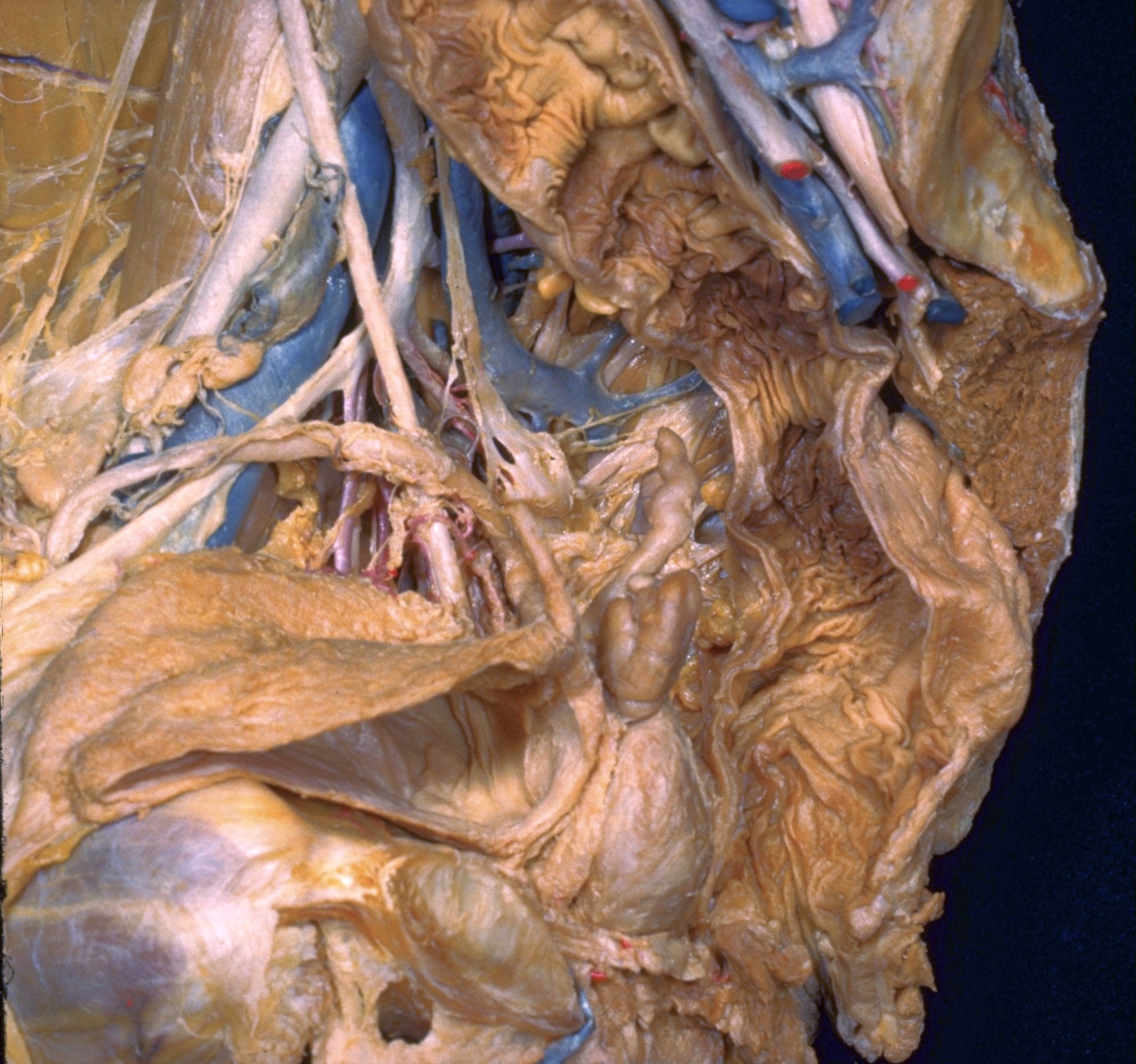
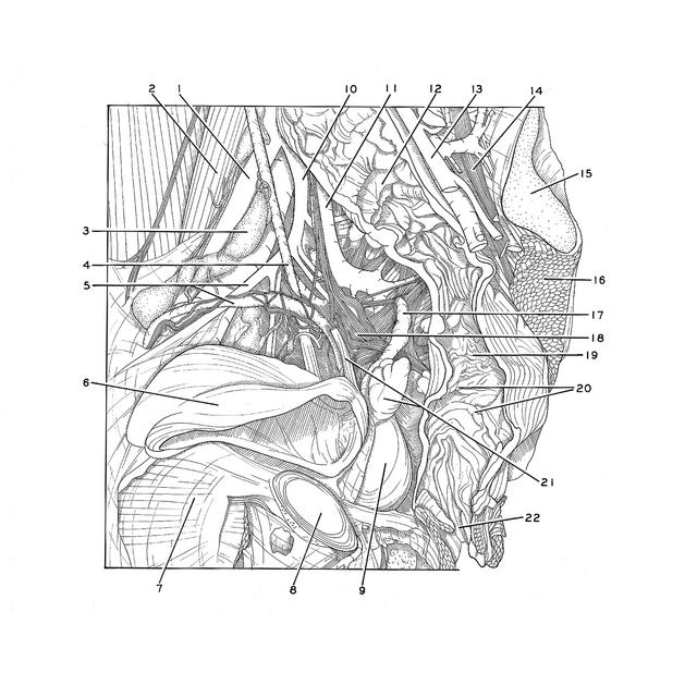
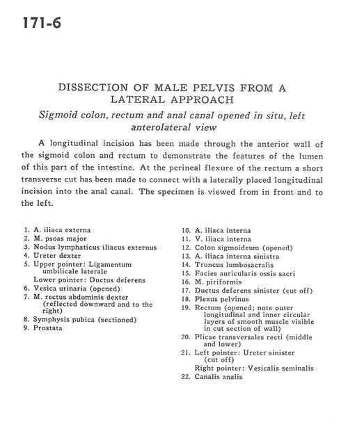
Dissection of male pelvis from a lateral approach
Sigmoid colon, rectum and anal canal opened in situ, left anterolateral view
Stanford holds the copyright to the David L. Bassett anatomical images and has assigned
Creative Commons license Attribution-Share Alike 4.0 International to all of the images.
For additional information regarding use and permissions,
please contact Dr. Drew Bourn at dbourn@stanford.edu.
Image #171-6
 |  | ||||||||||||||||||||||||||||||||||||||||||||||||
 |
|