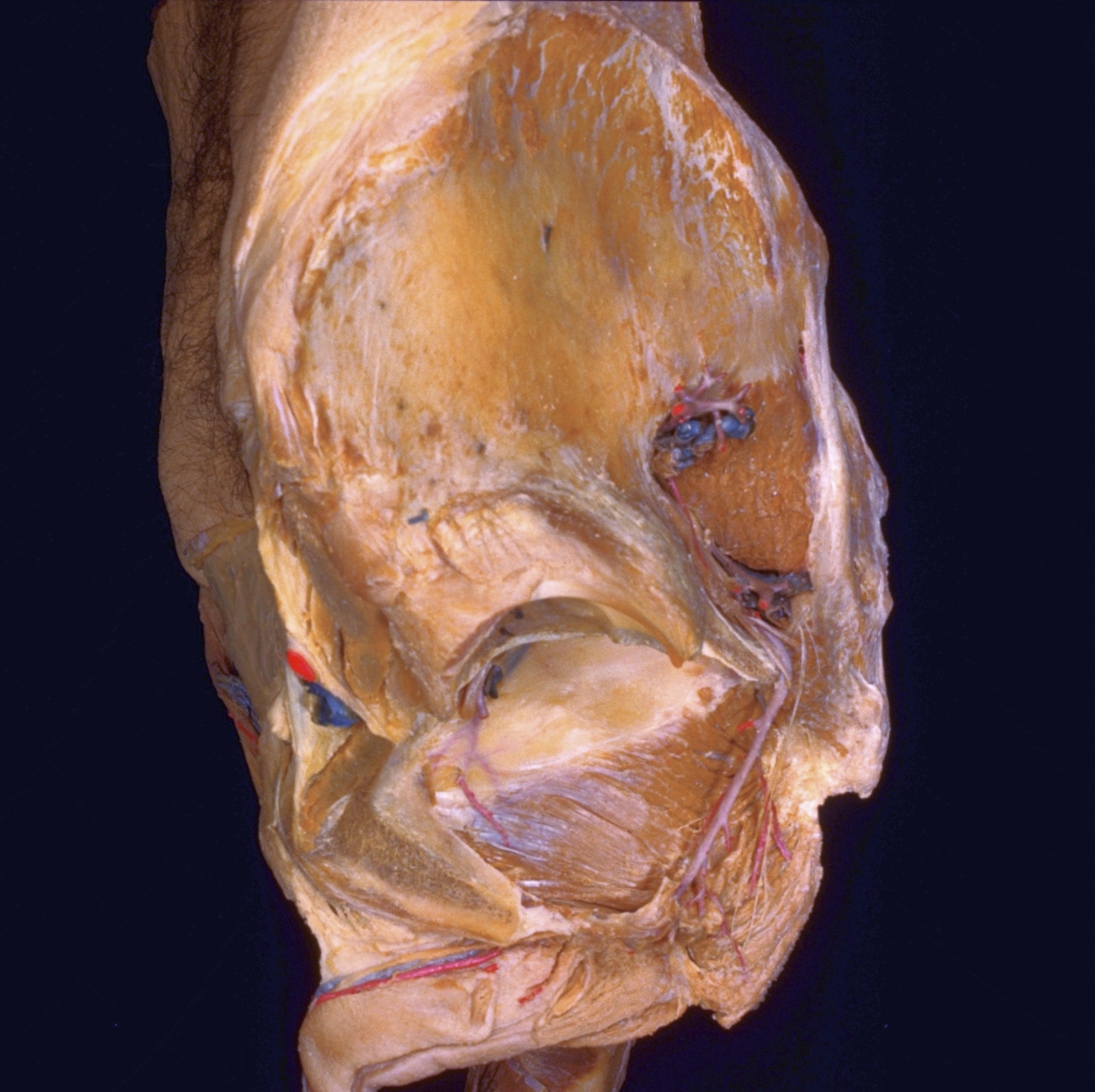
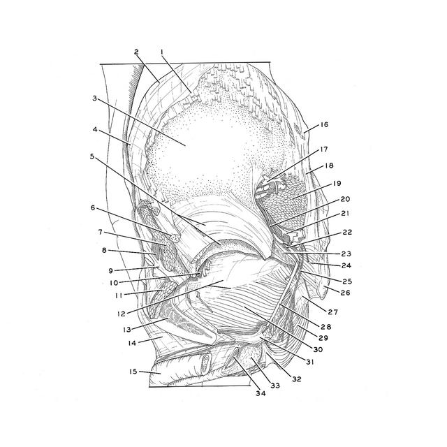
Left lateral view of pelvis with pelvic diaphragm exposed
For permissions information regarding the use of these images,
please contact Drew Bourn at dbourn@stanford.edu
Image #168-7
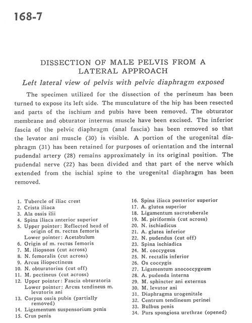
Dissection of male pelvis from a lateral approach
Left lateral view of pelvis with pelvic diaphragm exposed
The specimen utilized for the dissection of the perineum has been turned to expose its left side. The musculature of the hip has been resected and parts of the ischium and pubis have been removed. The obturator membrane and obturator internus muscle have been excised. The inferior fascia of the pelvic diaphragm (anal fascia) has been removed so that the levator ani muscle (30) is visible. A portion of the urogenital diaphragm (31) has been retained for purposes of orientation and the internal pudendal artery (28) remains approximately in its original position. The pudendal nerve (22) has been divided and that part of the nerve which extended from the ischial spine to the urogenital diaphragm has been removed.
1
.
Tubercle of iliac crest
2
.
Iliac crest
3
.
Ala of ilium
4
.
Anterior superior iliac spine
5
.
Upper pointer: Reflected head origin of rectus femoris muscle Lower pointer: Acetabulum
6
.
Origin of rectus femoris muscle
7
.
Iliopsoas muscle (cut across)
8
.
Femoral nerve (cut across)
9
.
Iliopectineal arch
10
.
Obturator nerve (cut off)
11
.
Pectineus muscle (cut across)
12
.
Upper pointer: Obturator fascia Lower pointer: Tendinous arch of levator ani muscle