X CLOSE

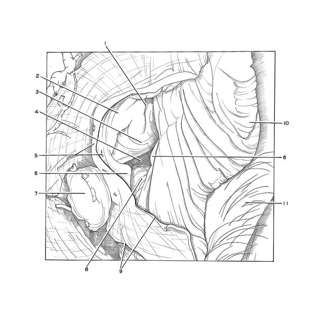
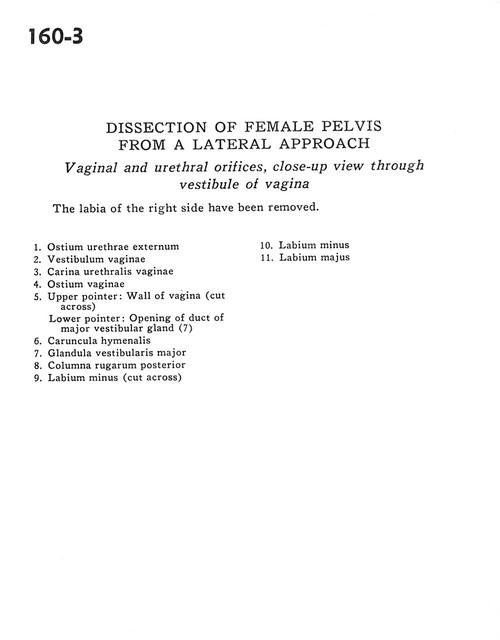
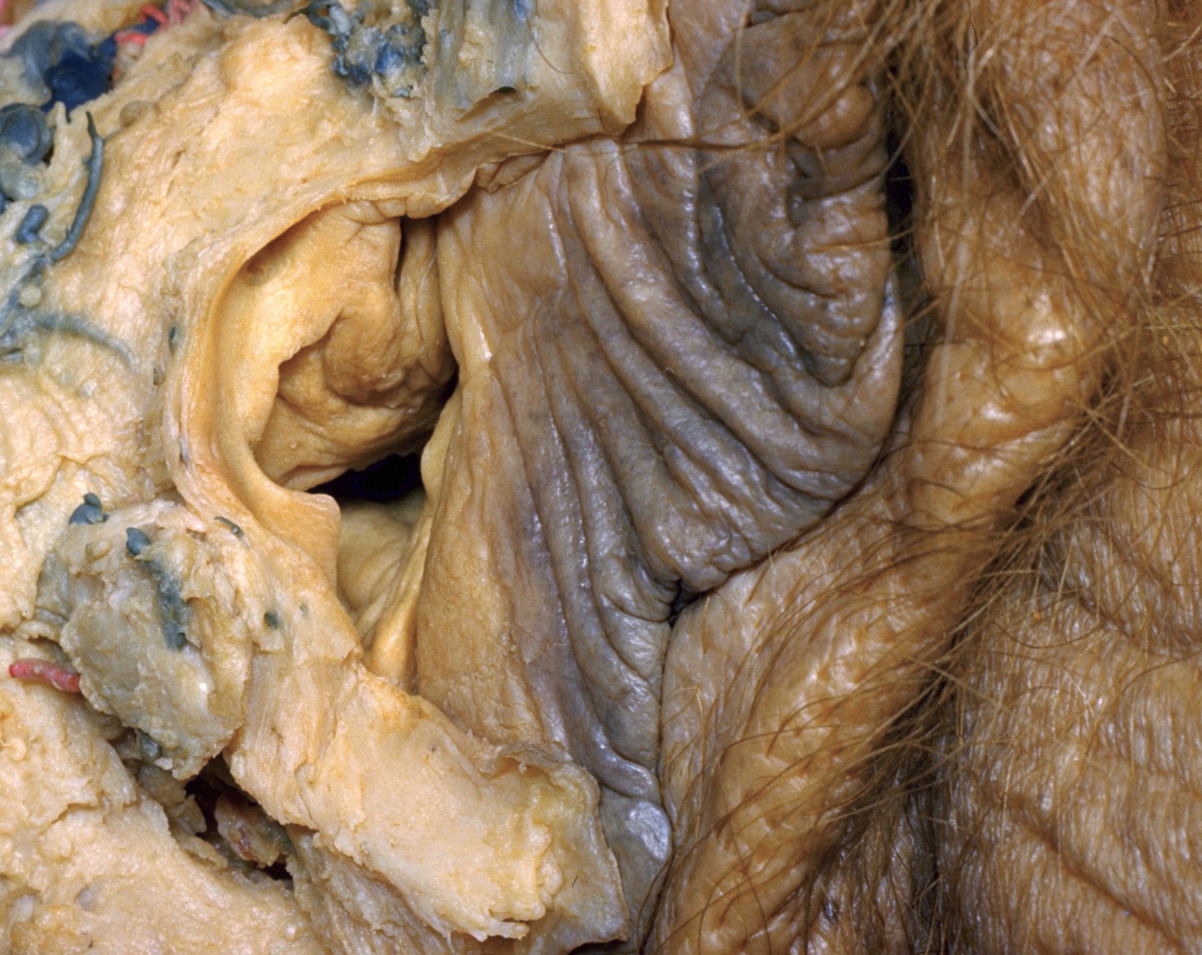
Dissection of female pelvis from a lateral approach

Vaginal and urethral orifices, close-up view through vestibule of vagina

Stanford holds the copyright to the David L. Bassett anatomical images and has assigned Creative Commons license Attribution-Share Alike 4.0 International to all of the images.
For additional information regarding use and permissions, please contact Dr. Drew Bourn at dbourn@stanford.edu.

Image #160-3
Bassett ImageDiagram Image
Legend Image
Dissection of female pelvis from a lateral approach
Vaginal and urethral orifices, close-up view through vestibule of vagina
The labia of the right side have been removed.
1 . External urethral opening
2 . Vestibule of vagina
3 . Urethral carina
4 . Vaginal opening
5 . Upper pointer: Wall of vagina (cut across) Lower pointer: Opening of duct of greater vestibular (Bartholin's) gland (7)
6 . Hymenal caruncle
7 . Greater vestibular (Bartholin's) gland
8 . Posterior neck fold
9 . Labium minus (cut across)
10 . Labium minus
11 . Labium majus