Dissection of female pelvis froma lateral approach
Superficial perineal space, close-up lateral view
For permissions information regarding the use of these images,
please contact Drew Bourn at dbourn@stanford.edu
Image #158-6
Dissection of female pelvis froma lateral approach
Superficial perineal space, close-up lateral view
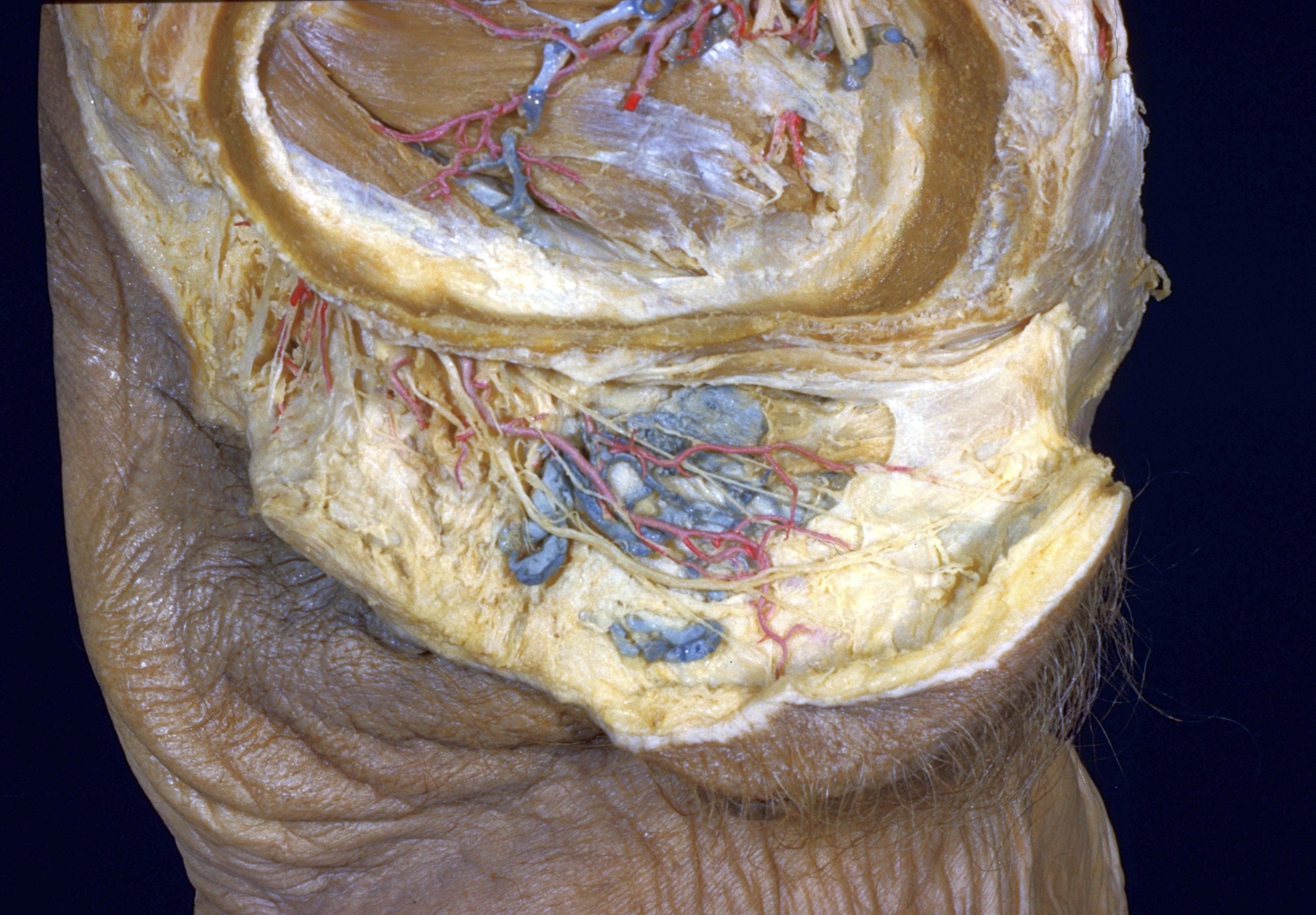
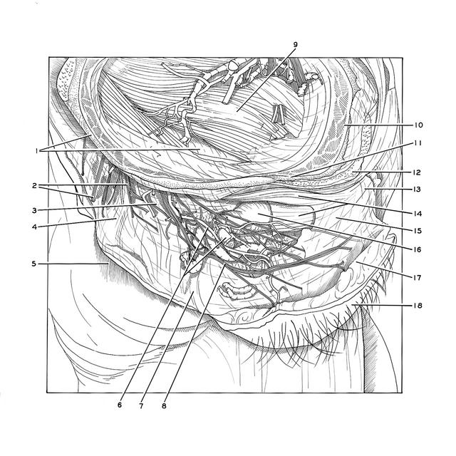
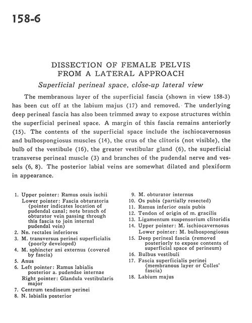
The membranous layer of the superficial fascia (shown in view 158-3) has been cut off at the labium majus (17) and removed. The underlying deep perineal fascia has also been trimmed away to expose structures within the superficial perineal space. A margin of the fascia remains anteriorly (15). The contents of the superficial space include the ischiocavernosus and bulbospongiosus muscles (14), the crus of the clitoris (not visible), the bulb of the vestibule (16), the greater vestibular gland (6), the superficial transverse perineal muscle (3) and branches of the pudendal nerve and vessels (6, 8). The posterior labial veins are somewhat dilated and plexiform in appearance.
1
.
Upper pointer: Ramus of ischium Lower pointer: Obturator fascia (pointer indicates location of pudendal canal, note branch of obturator vein passing through this fascia to join internal pudendal vein)