Dissection of female pelvis from a lateral approach
Superficial perineal space; obturator internus muscle; contents of obturator canal
For permissions information regarding the use of these images,
please contact Drew Bourn at dbourn@stanford.edu
Image #158-5
Dissection of female pelvis from a lateral approach
Superficial perineal space; obturator internus muscle; contents of obturator canal
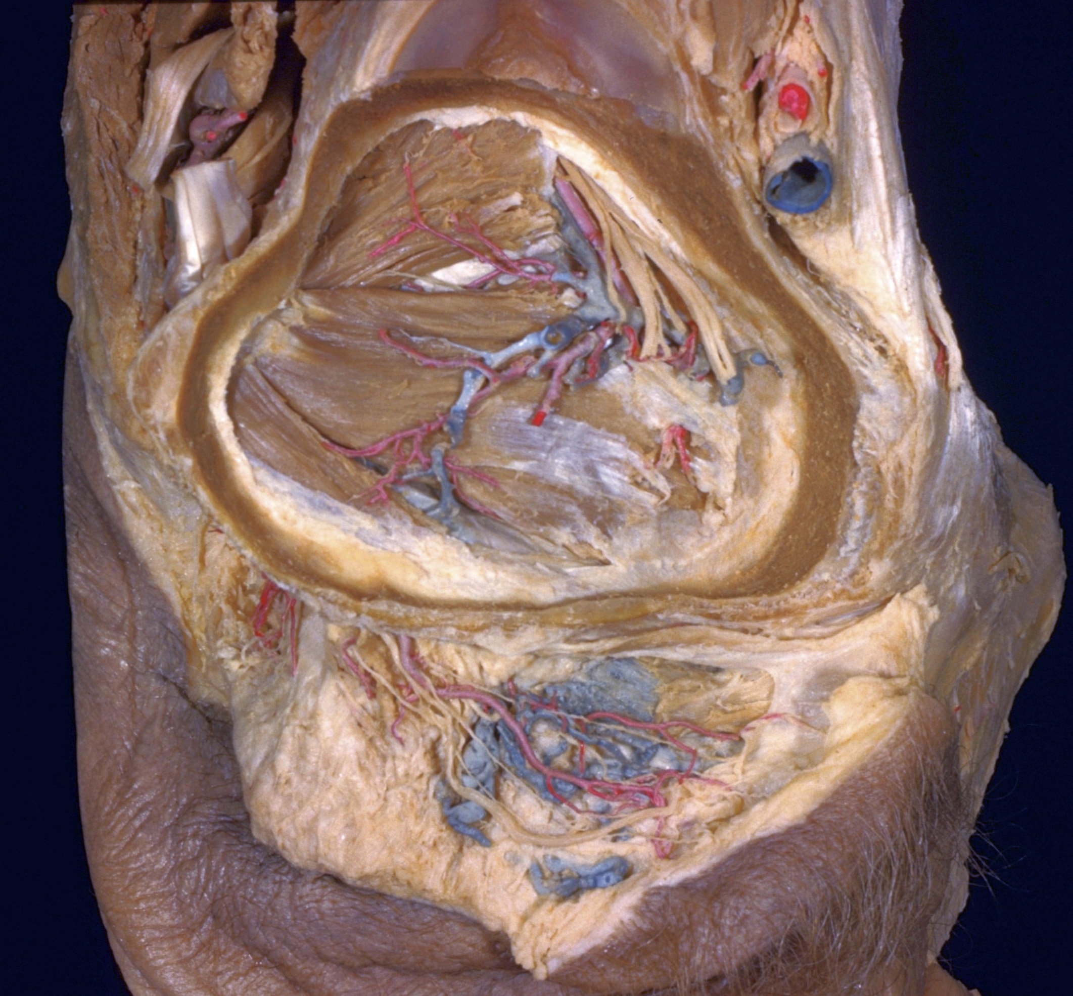
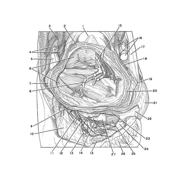
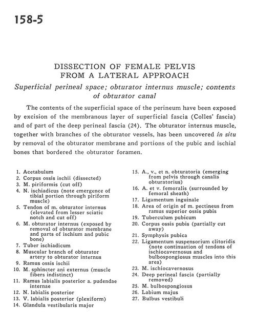
The contents of the superficial space of the perineum have been exposed by excision of the membranous layer of superficial fascia (Colles' fascia) and of part of the deep perineal fascia (24). The obturator internus muscle, together with branches of the obturator vessels, has been uncovered in situ by removal of the obturator membrane and portions of the pubic and ischial bones that bordered the obturator foramen.
1
.
Acetabulum
2
.
Body of ischium (dissected)
3
.
Piriform muscle (cut off)
4
.
Sciatic nerve (note emergence of tibial portion through piriform muscle)
5
.
Tendon of obturator internus muscle (elevated from lesser sciatic notch and cut off)
6
.
Obturator internus muscle (exposed by removal of obturator membrane and parts of ischium and pubic bone)
7
.
Ischial tuberosity
8
.
Muscular branch of obturator artery to obturator internus