X CLOSE

Female perineum

Deep perineal fascia

Stanford holds the copyright to the David L. Bassett anatomical images and has assigned Creative Commons license Attribution-Share Alike 4.0 International to all of the images.
For additional information regarding use and permissions, please contact Dr. Drew Bourn at dbourn@stanford.edu.

Image #157-1
Bassett ImageDiagram Image
Legend Image
Female perineum
Deep perineal fascia
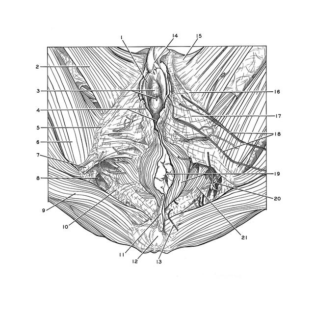
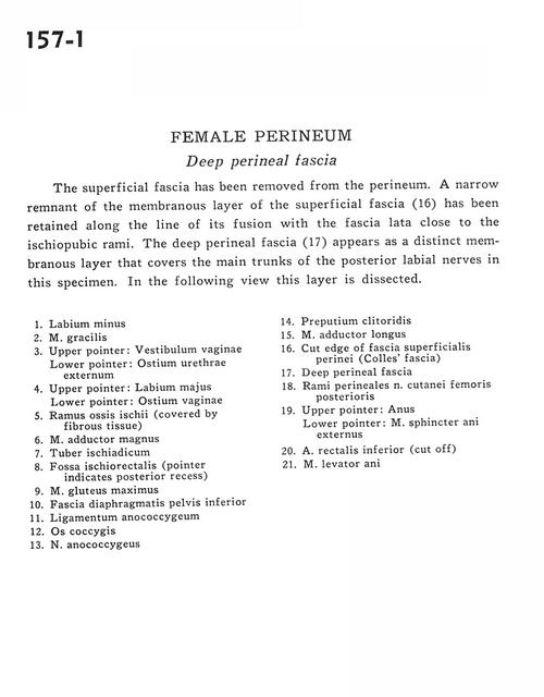
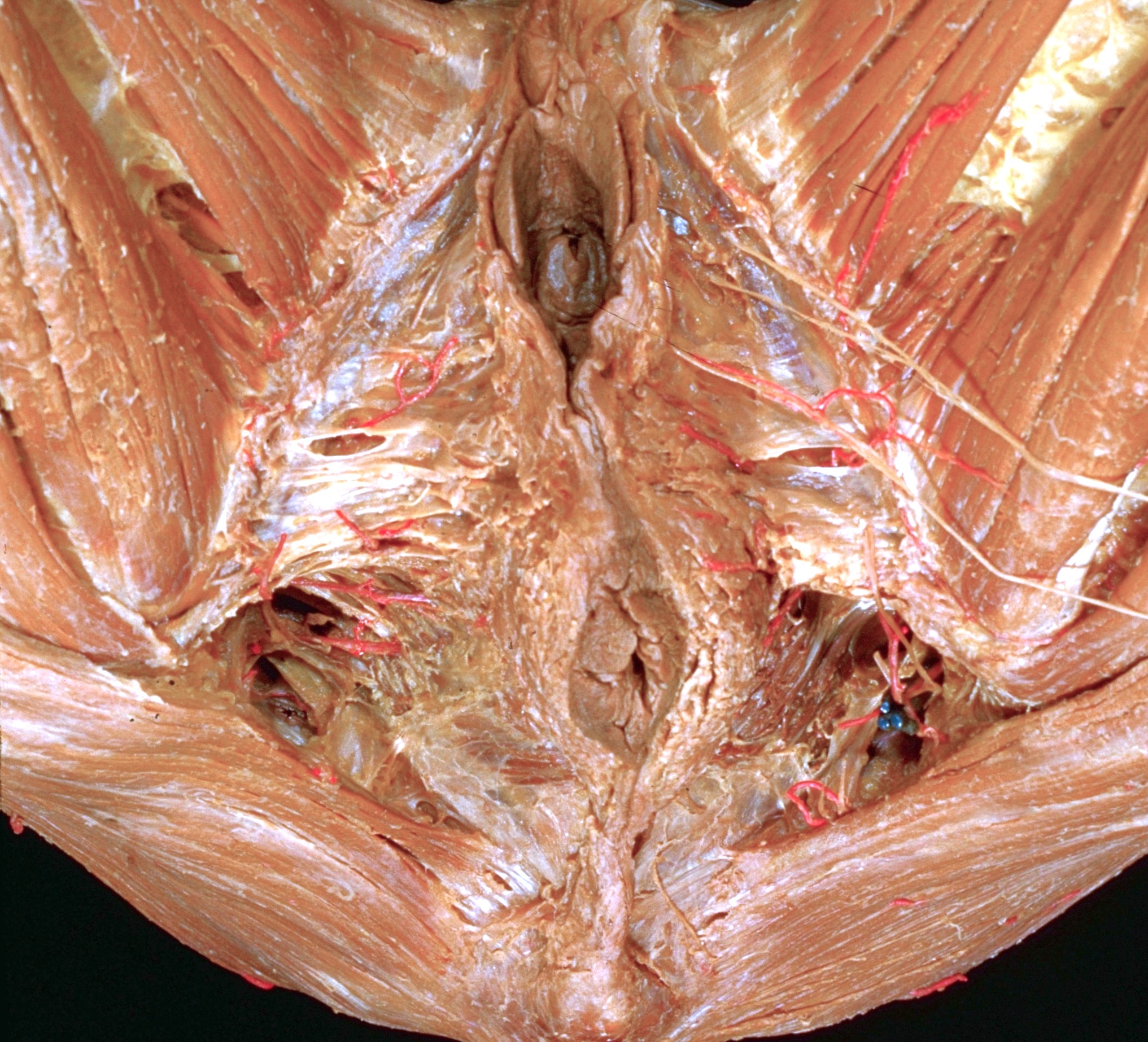
The superficial fascia has been removed from the perineum. A narrow remnant of the membranous layer of the superficial fascia (16) has been retained along the line of its fusion with the fascia lata close to the ischiopubic rami. The deep perineal fascia (17) appears as a distinct membranous layer that covers the main trunks of the posterior labial nerves in this specimen. In the following view this layer is dissected.
1 . Labium minus
2 . Gracilis muscle
3 . Upper pointer: Vestibule of vagina Lower pointer: External urethral opening
4 . Upper pointer: Labium majus Lower pointer: Vaginal opening
5 . Ramus of ischium (covered by fibrous tissue)
6 . Adductor magnus muscle
7 . Ischial tuberosity
8 . Ischiorectal fossa (pointer indicates posterior recess)
9 . Gluteus maximus muscle
10 . Inferior fascia of pelvic diaphragm
11 . Anococcygeal ligament
12 . Coccyx
13 . Anococcygeal nerve
14 . Prepuce of clitoris
15 . Adductor longus muscle
16 . Cut edge of superficial perineal fascia (Colles' fascia)
17 . Deep perineal fascia
18 . posterior perineal branches femoral cutaneous nerve
19 . Upper pointer: Anus Lower pointer: External anal sphincter muscle
20 . Inferior rectal artery (cut off)
21 . Levator ani muscle