X CLOSE

Ligaments and joints of lumbosacral spine and pelvic girdle

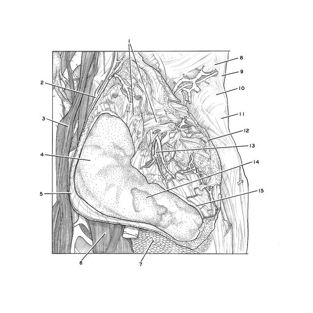
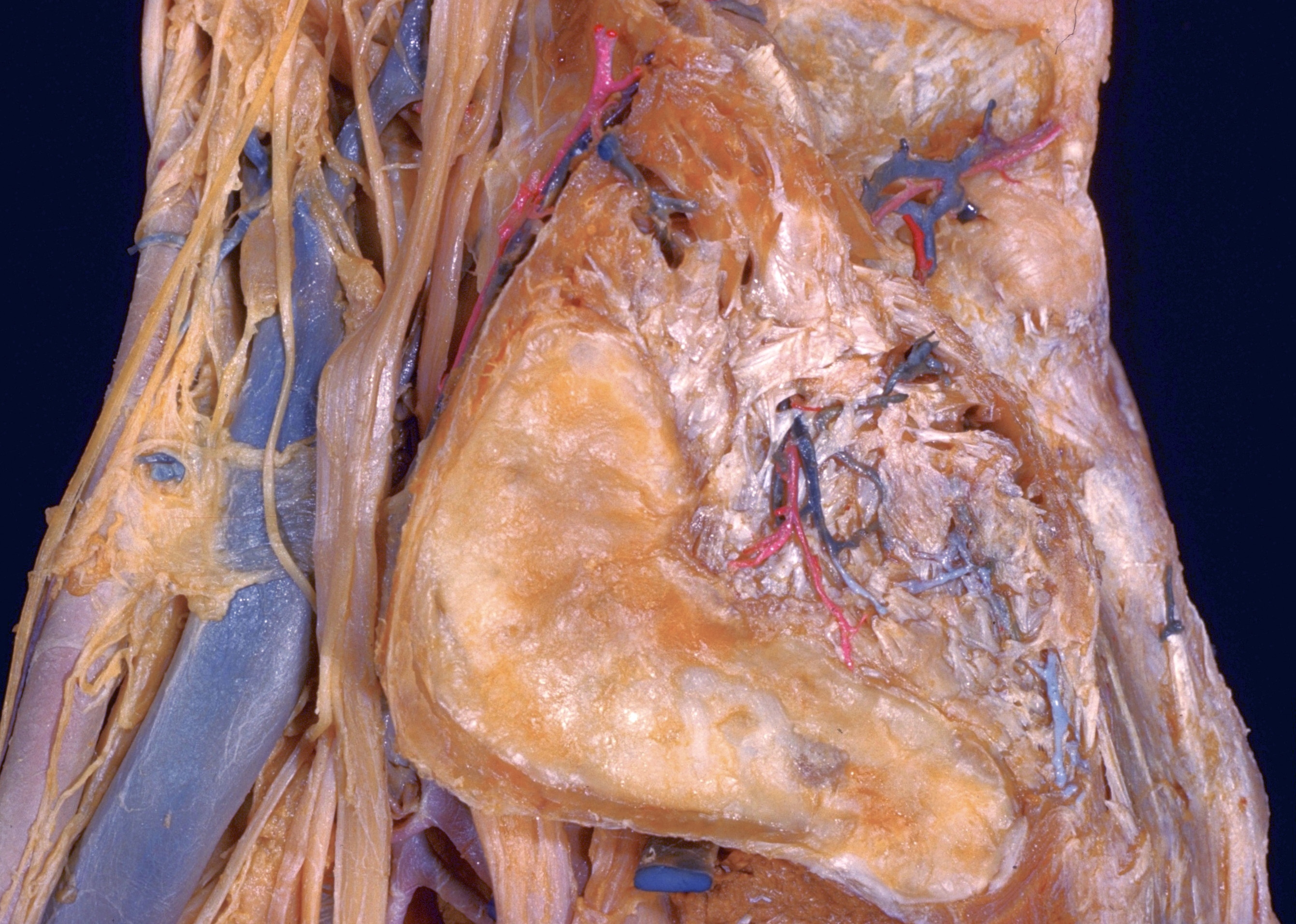
Sacroiliac joint opened; interosseous sacroiliac ligament, left lateral view

Stanford holds the copyright to the David L. Bassett anatomical images and has assigned Creative Commons license Attribution-Share Alike 4.0 International to all of the images.
For additional information regarding use and permissions, please contact Dr. Drew Bourn at dbourn@stanford.edu.

Image #156-2
Bassett ImageDiagram Image
Legend Image
Ligaments and joints of lumbosacral spine and pelvic girdle
Sacroiliac joint opened; interosseous sacroiliac ligament, left lateral view
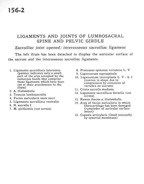
The left ilium has been detached to display the auricular surface of the sacrum and the interosseous sacroiliac ligaments.
1 . Interosseous sacroiliac ligament (pointer indicates only a small part of the area occupied by the numerous bands that comprise these ligaments which have been cut at their attachments to the ilium)
2 . Iliolumbar artery
3 . Lumbosacral trunk
4 . Articular surface of sacrum
5 . Ventral sacroiliac ligament
6 . Sacral nerve I
7 . Piriform muscle (cut across)
8 . Spinous process vertebra L. V
9 . Supraspinous ligament
10 . Interspinous ligament L. V - S. I (convex in shape due to compression by extension of vertebra on sacrum)
11 . Middle sacral crest
12 . Dorsal sacroiliac ligament (cut across)
13 . Iliac branch of iliolumbar artery
14 . Area of articular surface in which fibrocartilage has been damaged (remainder of auricular surface intact)
15 . Joint capsule (lined internally by synovial membrane)