Kidneys, suprarenal glands and posterior abdominal vessels, nerves and muscles
Blood supply and nerves of right suprarenal gland, close-up view
For permissions information regarding the use of these images,
please contact Drew Bourn at dbourn@stanford.edu
Image #150-3
Kidneys, suprarenal glands and posterior abdominal vessels, nerves and muscles
Blood supply and nerves of right suprarenal gland, close-up view
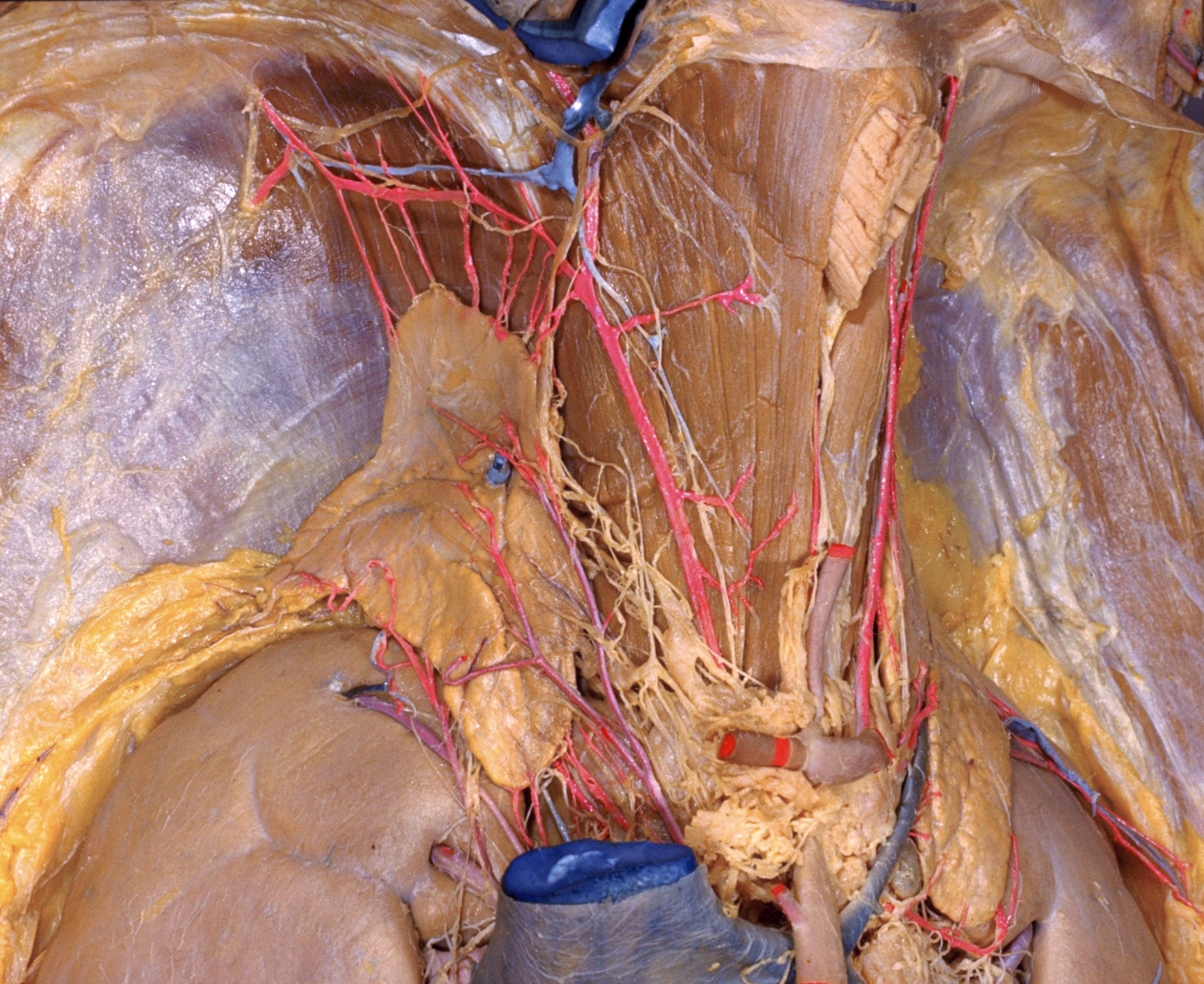
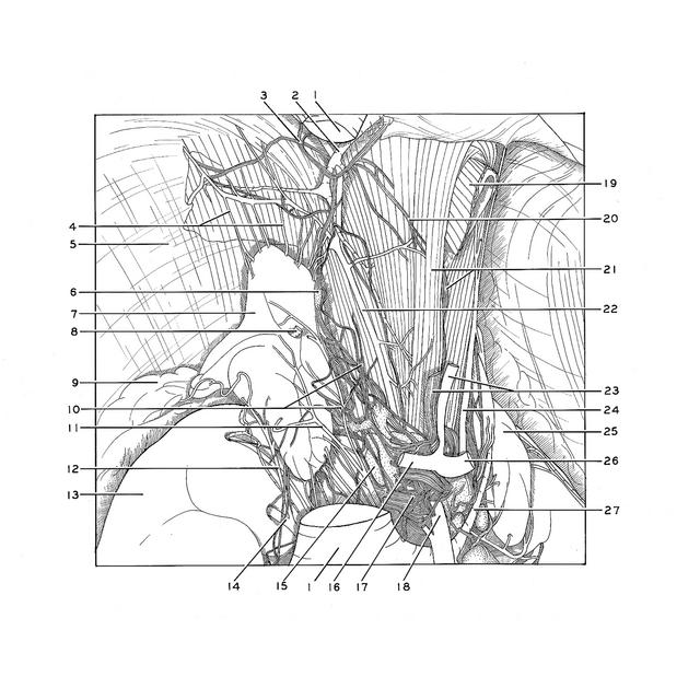
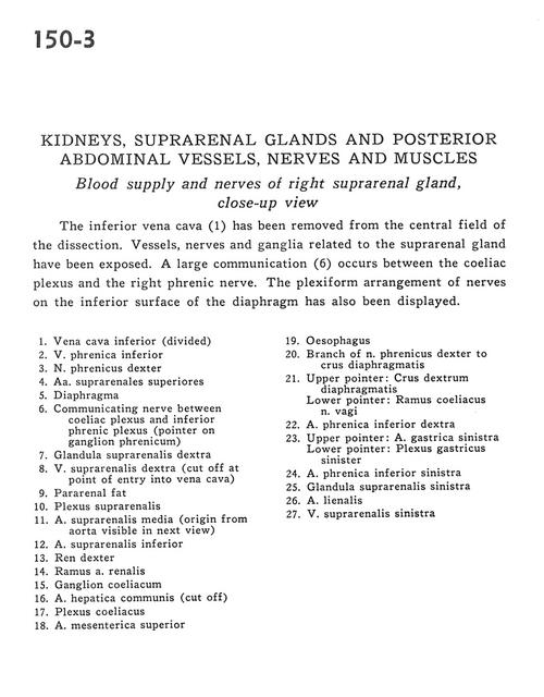
The inferior vena cava (1) has been removed from the central field of the dissection. Vessels, nerves and ganglia related to the suprarenal glands have been exposed. A large communication (6) occurs between the coeliac plexus and the right phrenic nerve. The plexiform arrangement of nerves on the inferior surface of the diaphragm has also been displayed.
1
.
Inferior vena cava (divided)
2
.
Inferior phrenic vein
3
.
Right phrenic nerve
4
.
Superior suprarenal arteries
5
.
Diaphragm
6
.
Communicating nerve between celiac plexus and inferior phrenic plexus (pointer on phrenic ganglion)
7
.
Right suprarenal gland
8
.
Right suprarenal vein (cut off at point of entry into vena cava)
9
.
Pararenal fat
10
.
Suprarenal plexus
11
.
Middle suprarenal artery (origin from aorta visible in next view)
12
.
Inferior suprarenal artery
13
.
Right kidney
14
.
Branch of renal artery
15
.
Celiac ganglion
16
.
Common hepatic artery (cut off)
17
.
Celiac plexus
18
.
Superior mesenteric artery
19
.
Esophagus
20
.
Branch of right phrenic nerve to crus of diaphragm
21
.
Upper pointer: Right crus of diaphragm Lower pointer: Celiac branch of vagus nerve
22
.
Right inferior phrenic artery
23
.
Upper pointer: Left gastric artery Lower pointer: Left gastric plexus