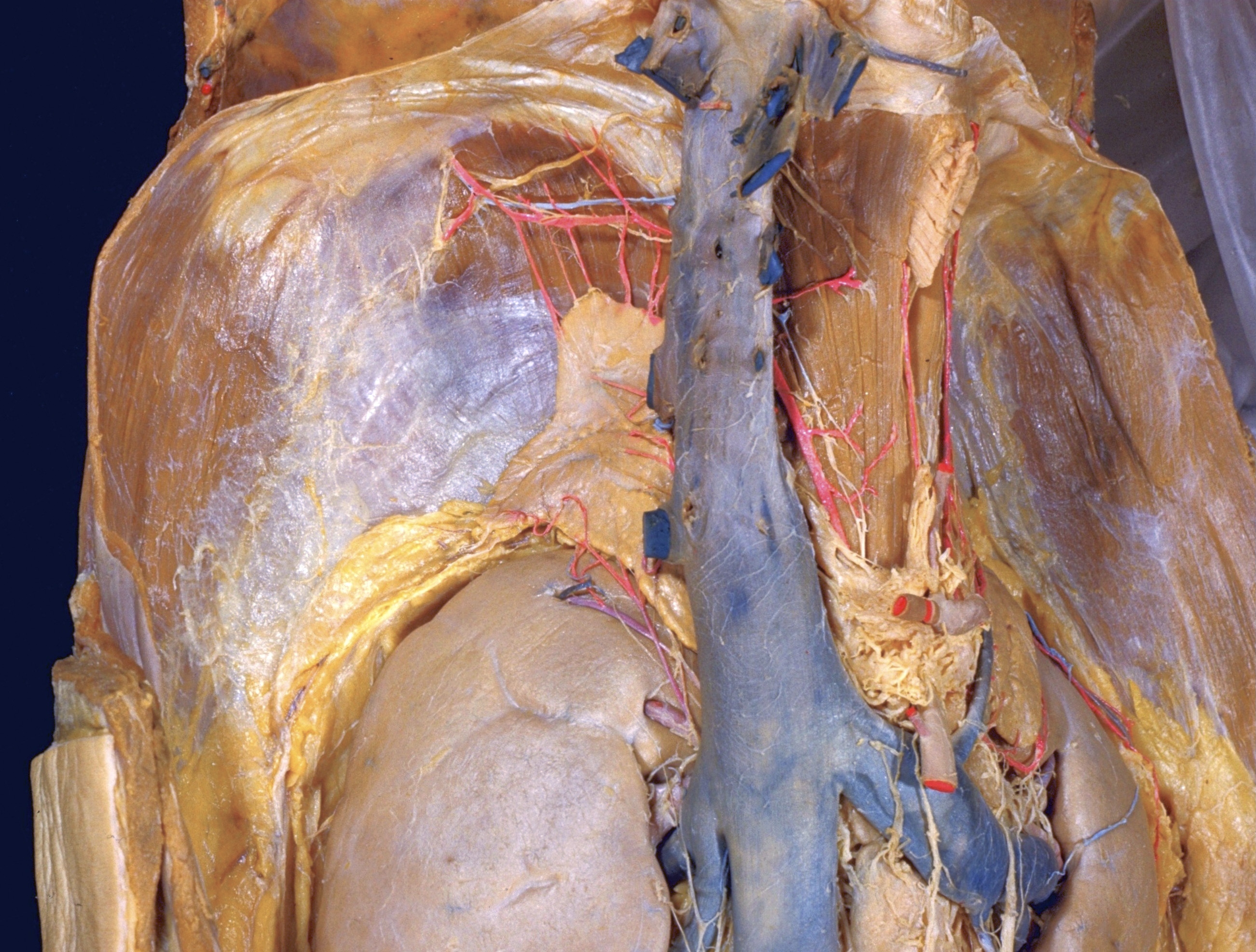
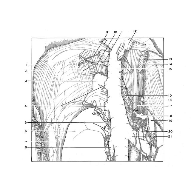
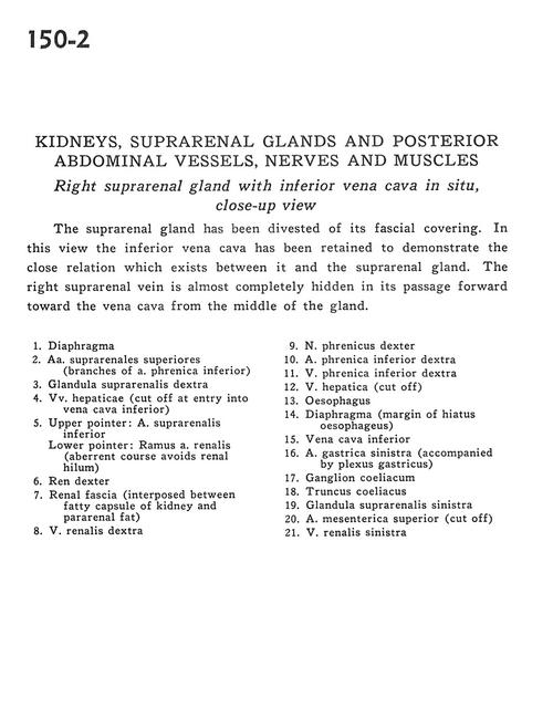
Kidneys, suprarenal glands and posterior abdominal vessels, nerves and muscles
Right suprarenal gland with inferior vena cava in situ, close-up view
Stanford holds the copyright to the David L. Bassett anatomical images and has assigned
Creative Commons license Attribution-Share Alike 4.0 International to all of the images.
For additional information regarding use and permissions,
please contact Dr. Drew Bourn at dbourn@stanford.edu.
Image #150-2
 |  | ||||||||||||||||||||||||||||||||||||||||||||||
 |
|