X CLOSE

Exploration of the brain from the medial aspect

Close-up view of central area of mid-sagittal section

Stanford holds the copyright to the David L. Bassett anatomical images and has assigned Creative Commons license Attribution-Share Alike 4.0 International to all of the images.
For additional information regarding use and permissions, please contact Dr. Drew Bourn at dbourn@stanford.edu.

Image #15-7
Bassett ImageDiagram Image
Legend Image
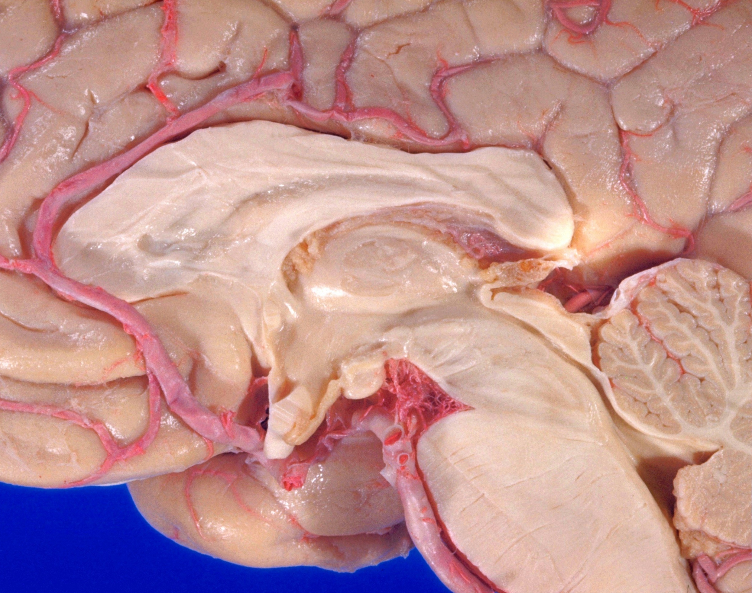
Exploration of the brain from the medial aspect
Close-up view of central area of mid-sagittal section
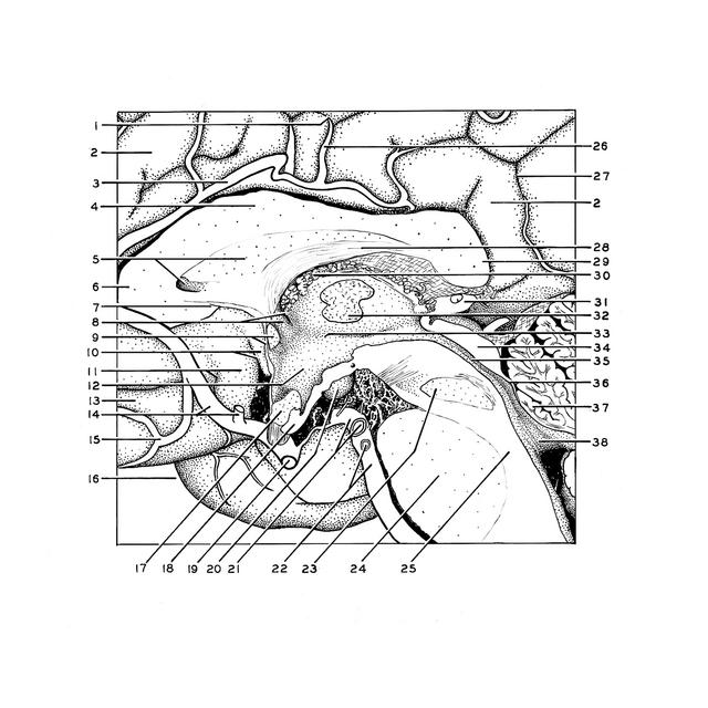
This view is centered on the diencephalon and third ventricle. The ventricular surfaces are indicated in the drawing in stipple.
1 . Subfrontal part cingulate sulci
2 . Cingulate gyrus
3 . Pericallosal artery
4 . Trunk corpus callosum
5 . Cavity of septum pellucidum and septum pellucidum
6 . Genu corpus callosum
7 . Rostrum of corpus callosum
8 . Free part of column of fornix and interventricular foramen
9 . Lamina terminalis and anterior commissure
10 . Posterior parolfactory sulcus and subcallosal gyrus (peduncles of corpus callosum)
11 . Anterior parolfactory sulcus and parolfactory area
12 . Hypothalamus
13 . Superior frontal gyrus
14 . Anterior cerebral artery and anterior communicating artery
15 . Orbital artery
16 . Temporal pole
17 . Optic chiasm
18 . Infundibulum of hypothalamus
19 . Internal carotid artery (cut off)
20 . Mamillary body
21 . Posterior cerebral artery (right and left) (cut off)
22 . Left basilar artery and superior cerebellar artery (cut off)
23 . Decussation brachium conjunctivum (superior cerebellar peduncle)
24 . Basilar part of pons
25 . Dorsal part of pons
26 . Middle medial frontal artery
27 . Subparietal sulcus
28 . Fornix (body)
29 . Corpus callosum (splenium)
30 . Choroid plexus third ventricle
31 . Pineal body
32 . Thalamus (pointer ends on cut surface of massa intermedia)
33 . Hypothalamic sulcus
34 . Quadrigeminal plate
35 . Cerebral aqueduct (Sylvian)
36 . Anterior medullary velum
37 . Cerebellar lingula
38 . Fourth ventricle