Kidneys, suprarenal, glands and posterior abdominal vessels, nerves and muscles
Longitudinal section of left kidney
Stanford holds the copyright to the David L. Bassett anatomical images and has assigned
Creative Commons license Attribution-Share Alike 4.0 International to all of the images.
For additional information regarding use and permissions,
please contact Dr. Drew Bourn at dbourn@stanford.edu.
Image #149-5
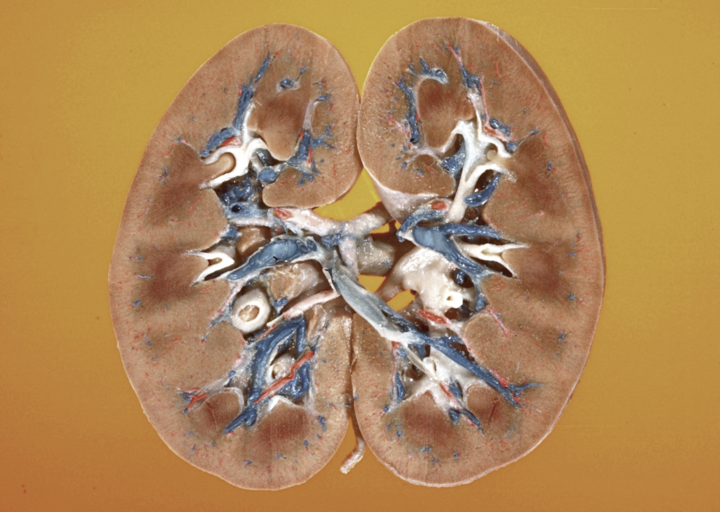 | 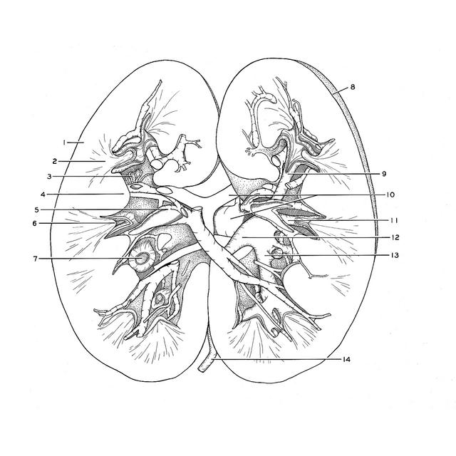 |
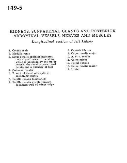 | | Kidneys, suprarenal, glands and posterior abdominal vessels, nerves and muscles | | Longitudinal section of left kidney | | | 1
.
| Cortex of kidney | | 2
.
| Medulla of kidney | | 3
.
| Renal sinus (pointer indicates only a small area of the sinus which is occupied by the major vessels, the renal calyces, renal pelvis, and a quantity of fat) | | 4
.
| Renal column | | 5
.
| Branch of renal vein split in sectioning kidney | | 6
.
| Renal papilla (sectioned) | | 7
.
| Renal papilla visible through sectioned wall of minor calyx | | 8
.
| Fibrous capsule | | 9
.
| Major renal calyx | | 10
.
| Renal artery and vein | | 11
.
| Minor calyx | | 12
.
| Renal pelvis | | 13
.
| Major renal calyx | | 14
.
| Ureter |
|
|


