Kidneys, suprarenal, glands and posterior abdominal vessels, nerves and muscles
Kidneys, ureters and suprarenal glands in situ, general view
Stanford holds the copyright to the David L. Bassett anatomical images and has assigned
Creative Commons license Attribution-Share Alike 4.0 International to all of the images.
For additional information regarding use and permissions,
please contact Dr. Drew Bourn at dbourn@stanford.edu.
Image #149-2
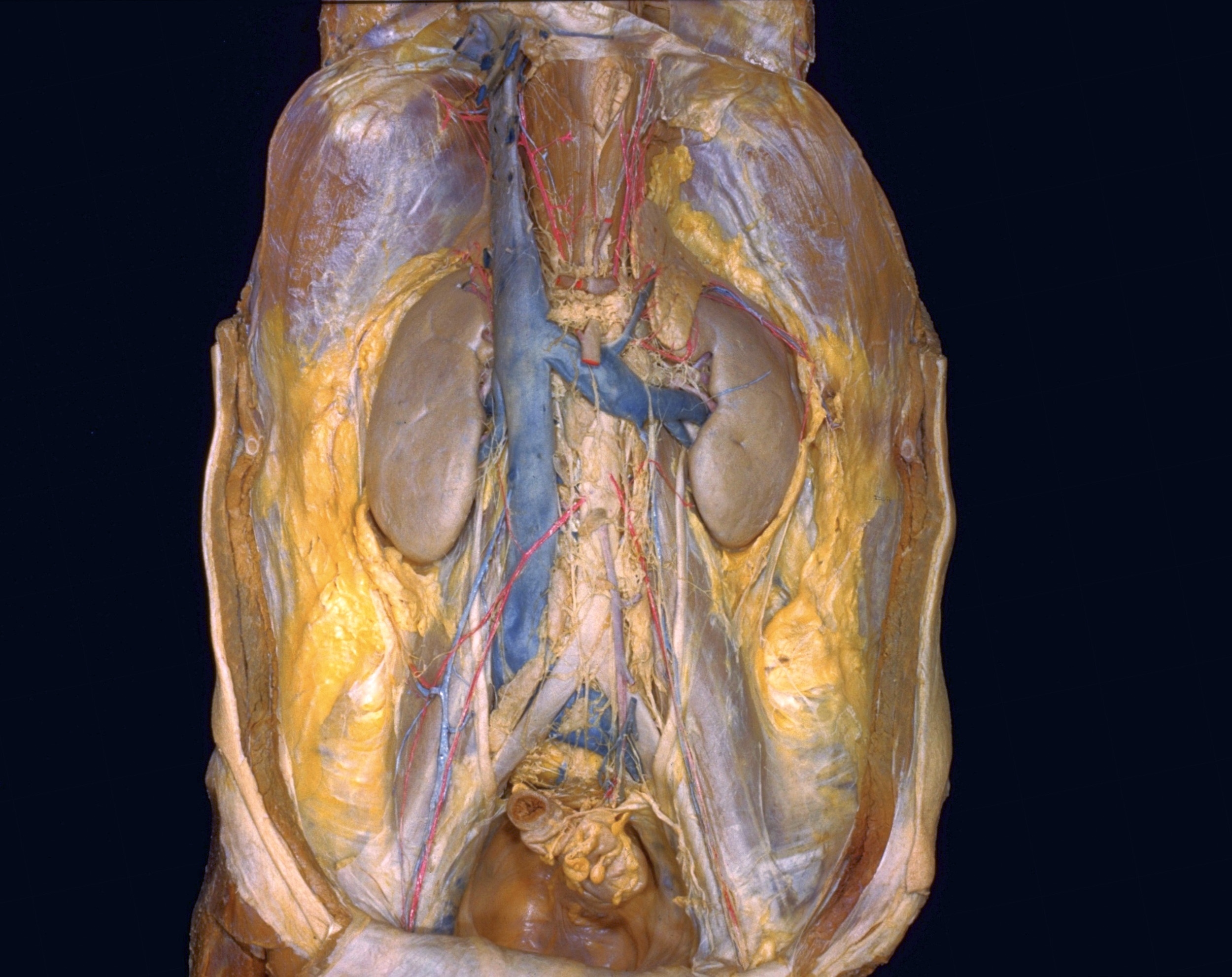 | 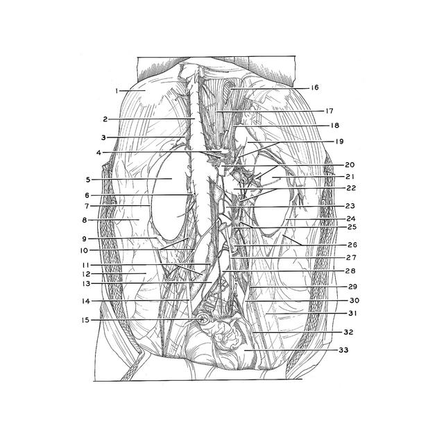 |
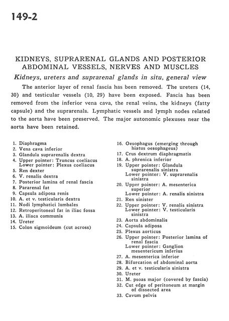 | | Kidneys, suprarenal, glands and posterior abdominal vessels, nerves and muscles | | Kidneys, ureters and suprarenal glands in situ, general view | | The anterior layer of renal fascia has been removed. The ureters (14, 30) and testicular vessels (10,29) have been exposed. Fascia has been removed from the inferior vena cava, the renal veins, the kidneys (fatty capsule) and the suprarenals. Lymphatic vessels and lymph nodes related to the aorta have been preserved. The major autonomic plexuses near the aorta have been retained. | | 1
.
| Diaphragm | | 2
.
| Inferior vena cava | | 3
.
| Right suprarenal gland | | 4
.
| Upper pointer: Celiac trunk Lower pointer: Celiac plexus | | 5
.
| Right kidney | | 6
.
| Right renal vein | | 7
.
| Posterior lamina of renal fascia | | 8
.
| Pararenal fat | | 9
.
| Fatty capsule of kidney | | 10
.
| Right testicular artery and vein | | 11
.
| Lumbar lymph nodes | | 12
.
| Retroperitoneal fat in iliac fossa | | 13
.
| Common iliac artery | | 14
.
| Ureter | | 15
.
| Sigmoid colon (cut across) | | 16
.
| Esophagus (emerging through esophageal hiatus) | | 17
.
| Right crus of diaphragm | | 18
.
| Inferior phrenic artery | | 19
.
| Upper pointer: Left suprarenal gland Lower pointer: Left suprarenal vein | | 20
.
| Upper pointer: Superior mesenteric artery Lower pointer: Left renal artery | | 21
.
| Left kidney | | 22
.
| Upper pointer: Left renal vein Lower pointer: Left testicular vein | | 23
.
| Abdominal aorta | | 24
.
| Fatty capsule | | 25
.
| Aortic plexus | | 26
.
| Upper pointer: Posterior layer of renal fascia Lower pointer: Inferior mesenteric ganglion | | 27
.
| Inferior mesenteric artery | | 28
.
| Bifurcation of abdominal aorta | | 29
.
| Left testicular artery and vein | | 30
.
| Ureter | | 31
.
| Psoas major muscle (covered by fascia) | | 32
.
| Cut edge of peritoneum at margin of dissected area | | 33
.
| Pelvic cavity |
|
|


