X CLOSE

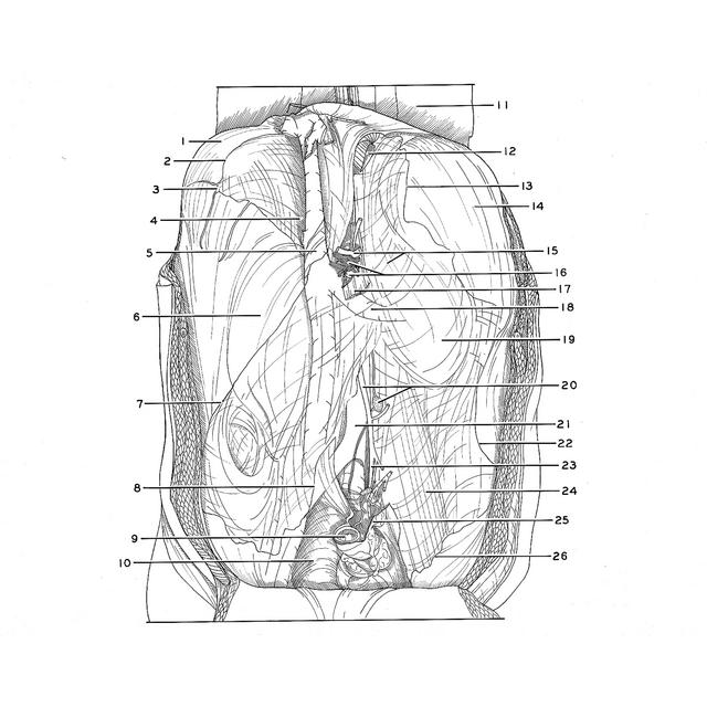
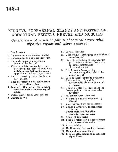
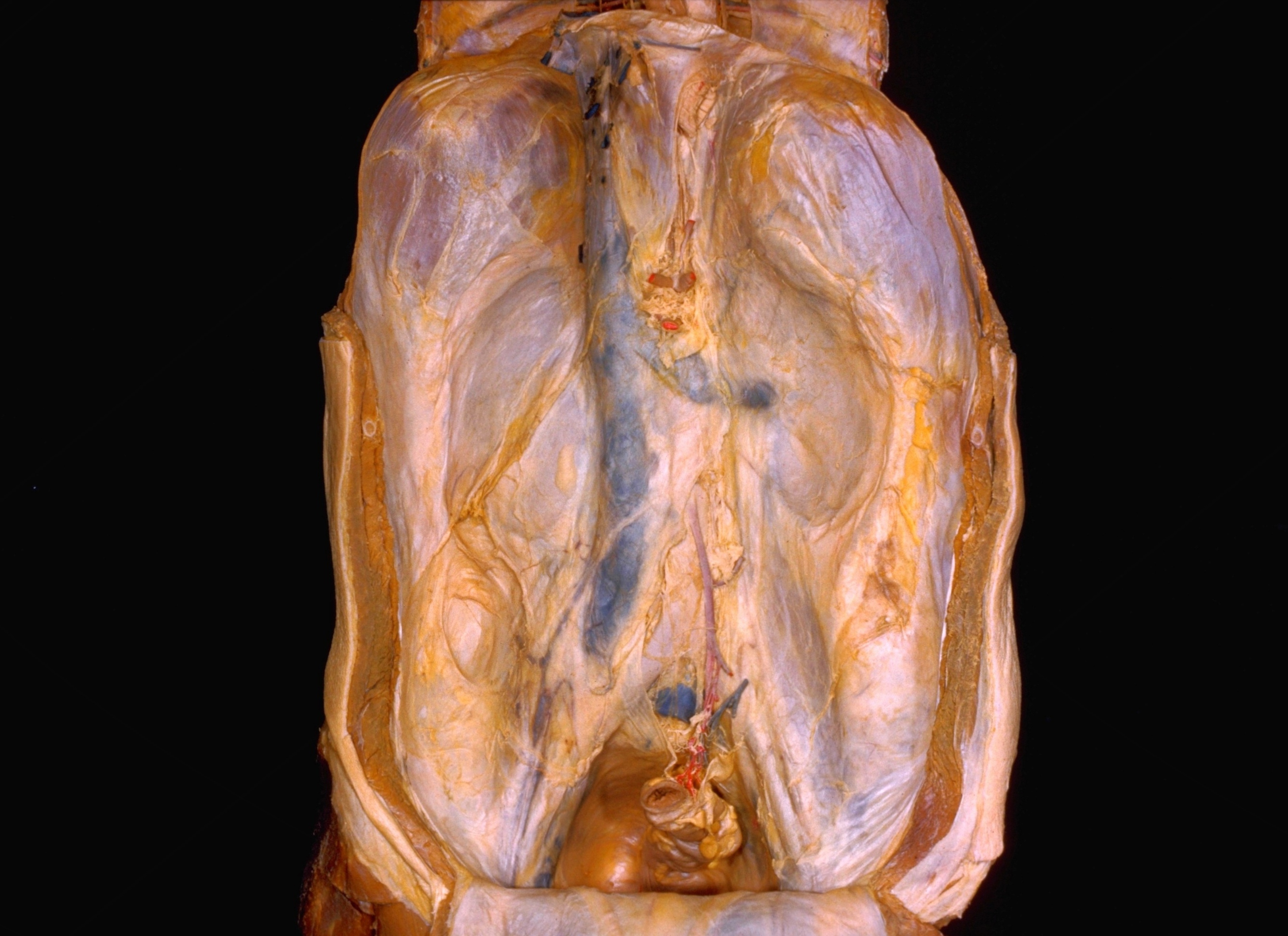
Kidneys, suprarenal glands and posterior abdominal vessels, nerves and muscles

General view of posterior part of abdominal cavity with digestive organs and spleen removed

Stanford holds the copyright to the David L. Bassett anatomical images and has assigned Creative Commons license Attribution-Share Alike 4.0 International to all of the images.
For additional information regarding use and permissions, please contact Dr. Drew Bourn at dbourn@stanford.edu.

Image #148-4
Bassett ImageDiagram Image
Legend Image
Kidneys, suprarenal glands and posterior abdominal vessels, nerves and muscles
General view of posterior part of abdominal cavity with digestive organs and spleen removed
1 . Diaphragm
2 . Coronary ligament of liver
3 . Right triangular ligament
4 . Right suprarenal gland (covered by fascia)
5 . Inferior vena cava (pointer on peritonealized part of vena cava which passed behind epiploic foramen in intact specimen)
6 . Kidney (covered by renal fascia and peritoneum)
7 . Line of reflection of peritoneum onto ascending colon
8 . Line of reflection of peritoneum onto left side of mesentery of ileum
9 . Sigmoid colon (cut across)
10 . Pelvic cavity
11 . Thoracic cavity
12 . Esophagus (emerging below esophageal hiatus)
13 . Line of reflection of gastrosplenic ligament (lower down this becomes phrenicolienal ligament)
14 . Diaphragm (covered by peritoneum against which the spleen rests)
15 . Left pointer: Celiac trunk Right pointer: Left suprarenal gland (covered by fascia)
16 . Upper pointer: Celiac plexus Lower pointer: Superior mesenteric artery
17 . Suspensory muscle of duodenum
18 . Left renal vein (covered by fascia)
19 . Kidney (covered by renal fascia)
20 . Upper pointer: Inferior mesenteric artery Lower pointer: Inferior mesenteric ganglion
21 . Abdominal aorta
22 . Line of reflection of peritoneum onto descending colon
23 . Sigmoid artery
24 . Iliopsoas muscle (covered by fascia)
25 . Mesosigmoid colon
26 . Line of attachment of mesosigmoid colon