Exploration of liver, gall bladder, pancreas, duodenum and spleen
Dissection of duodenum (continued).
Stanford holds the copyright to the David L. Bassett anatomical images and has assigned
Creative Commons license Attribution-Share Alike 4.0 International to all of the images.
For additional information regarding use and permissions,
please contact Dr. Drew Bourn at dbourn@stanford.edu.
Image #147-5
 | 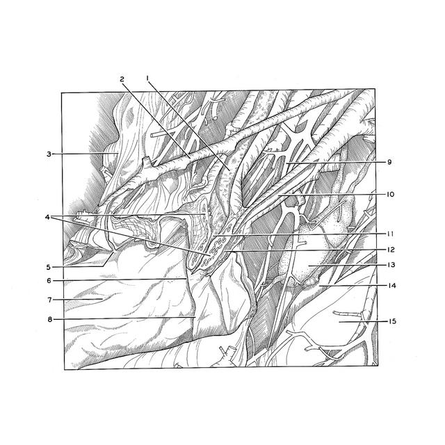 |
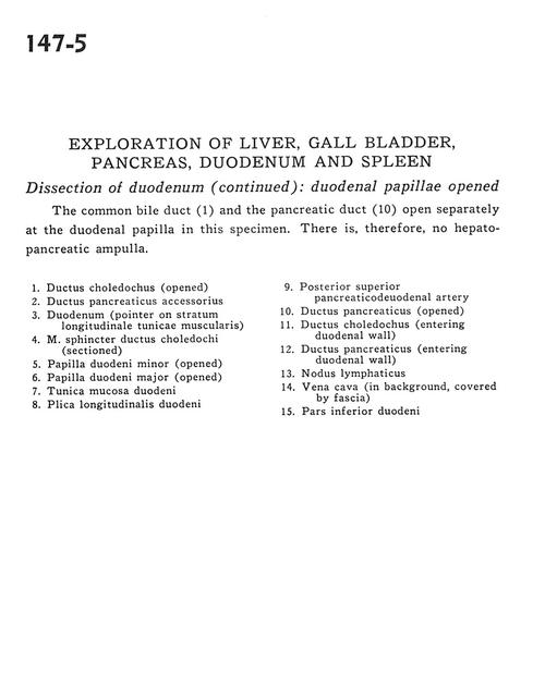 | | Exploration of liver, gall bladder, pancreas, duodenum and spleen | | Dissection of duodenum (continued). | | The common bile duct (1) and the pancreatic duct (10) open separately at the duodenal papilla in this specimen. There is, therefore, no hepatopancreatic ampulla. | | 1
.
| Common bile duct (opened) | | 2
.
| Accessory pancreatic duct | | 3
.
| Duodenum (pointer on longitudinal muscle layer of stomach) | | 4
.
| Sphincter muscle of common bile duct (sectioned) | | 5
.
| Minor duodenal papilla (opened) | | 6
.
| Major duodenal papilla (opened) | | 7
.
| Muscular layer of duodenum | | 8
.
| Longitudinal duodenal fold | | 9
.
| Posterior superior pancreaticoduodenal artery | | 10
.
| Pancreatic duct (opened) | | 11
.
| Common bile duct (entering duodenal wall) | | 12
.
| Pancreatic duct (entering duodenal wall) | | 13
.
| Lymph node | | 14
.
| Vena cava (in background, covered by fascia) | | 15
.
| Inferior part of duodenum |
|
|


